Extensor retinaculum of hand (Latin: retinaculum musculorum extensorum (membri superioris))
Also called: extensor retinaculum of wrist; dorsal carpal ligament
The extensor retinaculum of the hand is:
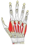 Back of the hand
 Forearm and hand (posterior view)
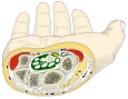 Cross-section of the wrist
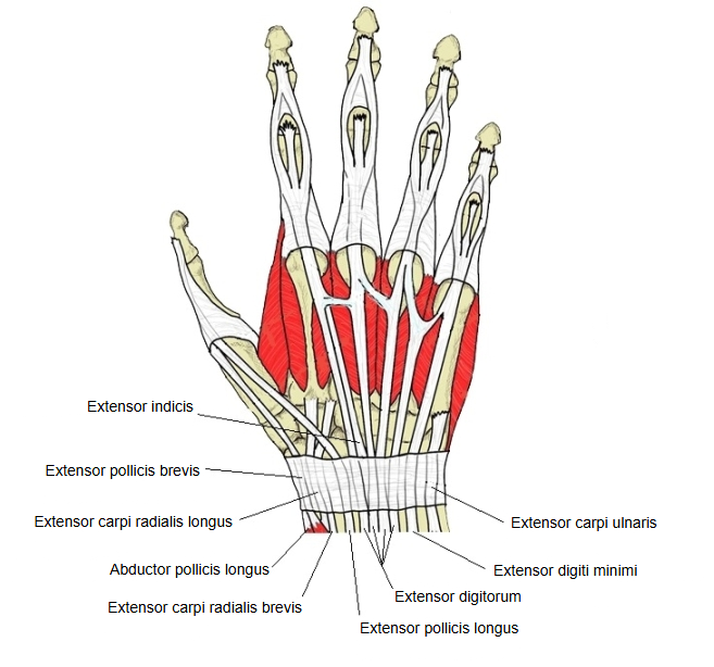
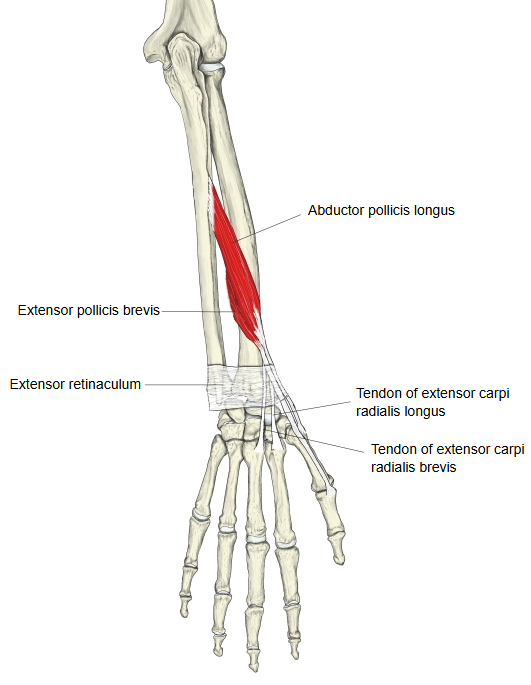
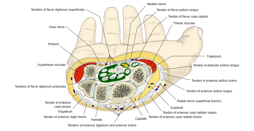
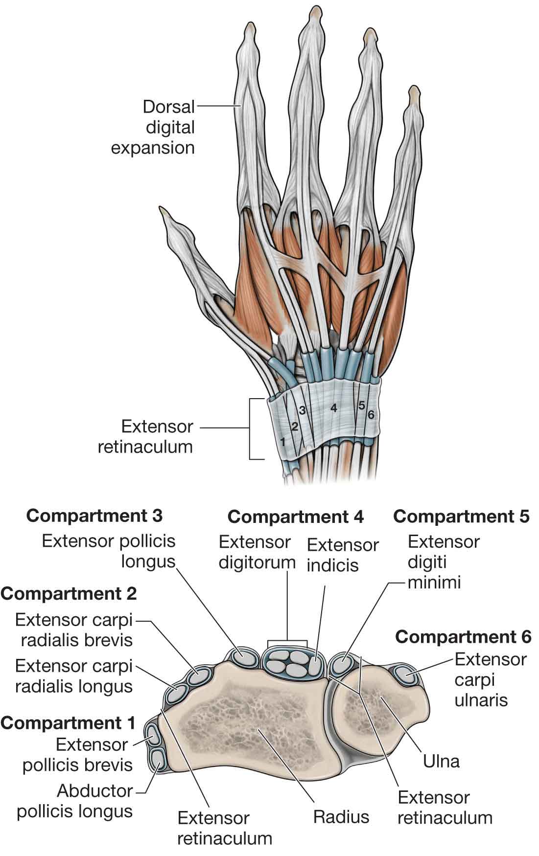
The main extensor tendons that pass beneath it are as follows (in order from the thumb side; the compartment number is given in brackets).
The retinaculum is a strong fibrous band, a thickened part of the deep fascia of the forearm reinforced by transverse fibres, which runs obliquely downwards and medially across the back of the wrist. Laterally it is attached to the lateral margin of the radius near the styloid process; medially it is attached not to the ulna, whose distance from the radius changes in pronation and supination, but to the triquetrum and pisiform. On its way it is also attached to the ridges on the back of the radius, including the dorsal tubercle, and so forms six compartments, each lined by a tendon sheath. On the front of the wrist the same fascia continues as the palmar carpal ligament, which lies superficial to the flexor retinaculum (it is not the same structure).
Reference websites
[Wikipedia]
· Extensor retinaculum of the hand, Wikipedia.
[Websites with illustrations]
· Back of the hand with the extensor retinaculum and the extensor tendons (Musculoskeletal Key)
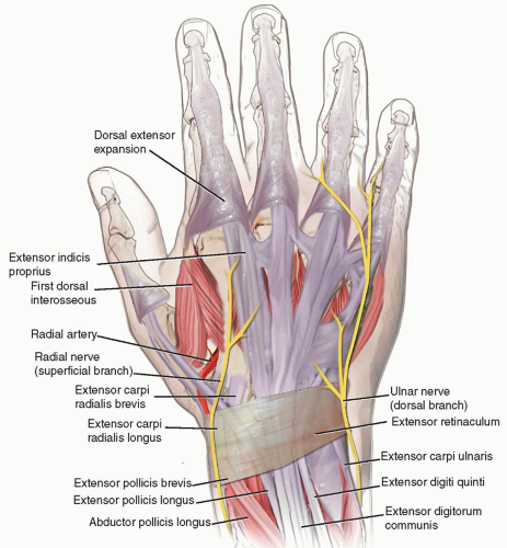
· Synovial sheaths of the extensor tendons under the retinaculum (Pinterest (image))

· The six extensor compartments, with a cross-section (Musculoskeletal Key)
· Extensor retinaculum seen from the radial side (DoctorLib)

Related terms
- Flexor retinaculum (hand)
- Superior extensor retinaculum
- Inferior extensor retinaculum
- Deep fascia
- Tendon sheath
- Radiocarpal joint
- Dorsal tubercle of radius
References and sources
- Further reading: Extensor retinaculum of the hand, Wikipedia.
- Federative International Programme for Anatomical Terminology (FIPAT). Terminologia Anatomica, 2nd ed. (TA2), 2019.