Middle pharyngeal constrictor (Latin: musculus constrictor pharyngis medius)
- Overview
- Illustrations
- Origin / insertion
- Action
- Nerve / blood supply
- Reference websites
- (Wikipedia, websites with illustrations)
- Related terms
Overview
- Superficial muscles of the neck (10)
- Deep muscles of the neck (8)
- Muscles of the pharynx (6)
- Pharyngeal constrictors (3) : superior, middle, inferior (its lowest part is the cricopharyngeus)
- Pharyngeal elevators (3) : stylopharyngeus, salpingopharyngeus, palatopharyngeus
· A fan-shaped muscle, smaller than the inferior constrictor. All three constrictors end behind in the midline on the pharyngeal raphe, overlapping like stacked cups.
- Longitudinal muscles : raise the pharynx: the pharyngeal elevators
- Circular muscles : narrow the pharynx: the pharyngeal constrictors
- Superior constrictor : the uppermost
- Middle constrictor : in the middle
- Inferior constrictor : the lowest; it has two parts (thyropharyngeus and cricopharyngeus)
Illustrations
More figures: Pharyngeal constrictors (lateral view) · Pharyngeal constrictors (posterior view) · Muscles of the neck (list)
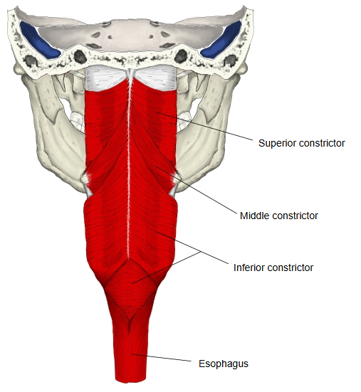
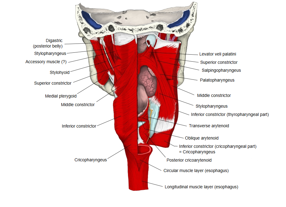
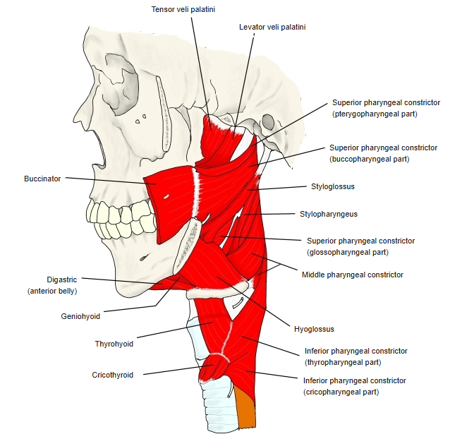
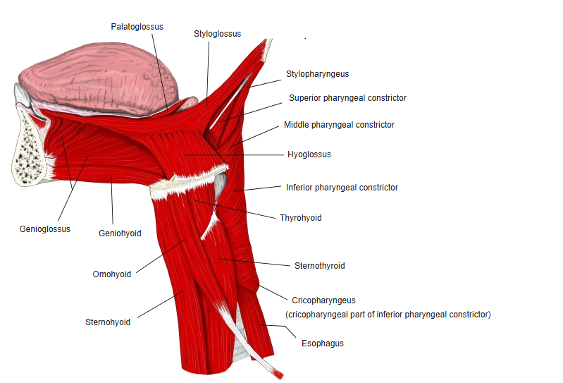
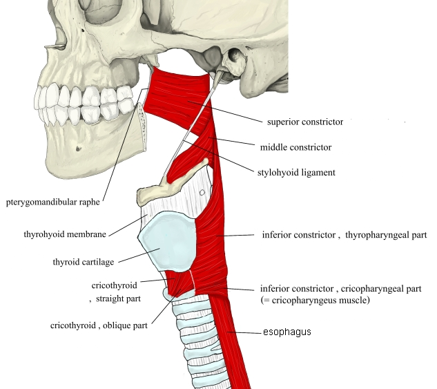
Origin / insertion
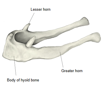
[Origin] : the hyoid bone: the lesser horn (chondropharyngeal part) and the whole upper border of the greater horn (ceratopharyngeal part), and the lower end of the stylohyoid ligament.
The fibres spread out from this narrow origin: the upper fibres climb and overlap the superior constrictor from outside, the middle fibres run horizontally, and the lower fibres descend under the inferior constrictor.
[Insertion] : the pharyngeal raphe in the midline at the back of the pharynx, where it meets the muscle of the other side.
 Hyoid bone
Action
Narrows the middle part of the pharynx. In swallowing, the three constrictors contract one after another from above downward and push the food into the esophagus.
Nerve / blood supply
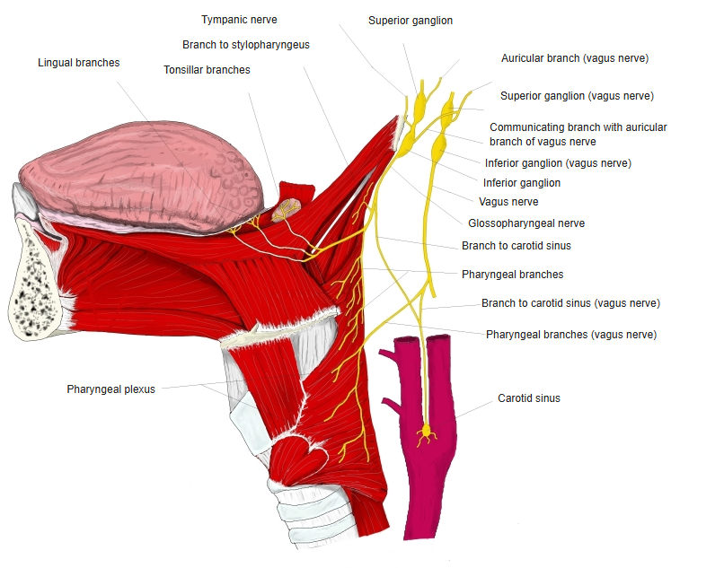
· Nerve : the pharyngeal plexus, through the pharyngeal branches of the vagus nerve (cranial nerve X).
· Arteries : mainly the ascending pharyngeal artery, with the tonsillar branch of the facial artery and branches of the lingual artery and superior thyroid artery.
· Veins : the pharyngeal venous plexus, draining by the pharyngeal veins to the internal jugular vein.
 Pharyngeal plexus
Reference websites
[Wikipedia]
· Middle pharyngeal constrictor muscle, Wikipedia.
[Websites with illustrations]
· Pharyngeal constrictors, lateral view (image; the superior constrictor highlighted, the middle constrictor below it) (Wikimedia Commons)

Related terms
- Constrictors of pharynx
- Pharyngeal raphe
- Greater horn
- Lesser horn
- Pharyngeal plexus
- Superior pharyngeal constrictor
- Inferior pharyngeal constrictor
- Hyoid bone
- Pharynx
References and sources
- FIPAT. Terminologia Anatomica, 2nd ed. (TA2), 2019: musculus constrictor pharyngis medius.
- Middle pharyngeal constrictor muscle, Wikipedia.