Latin: os cuboideum
Overview
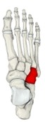
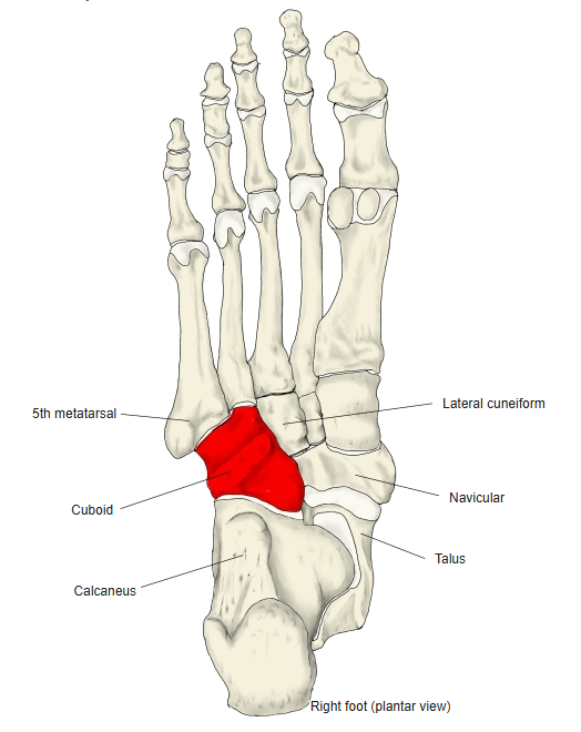
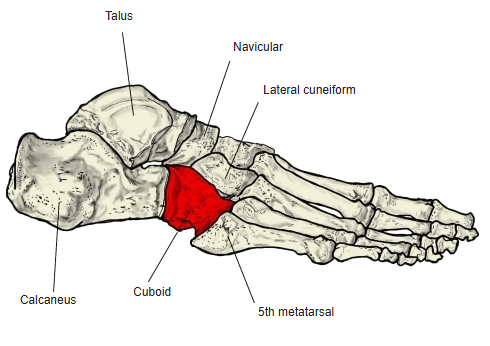
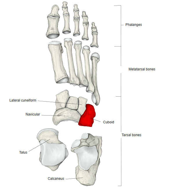
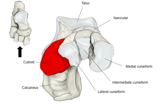
- One of the seven tarsal bones; it is the most lateral of the distal row of tarsal bones.
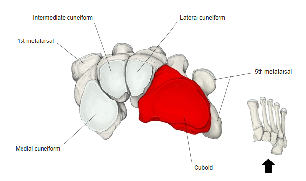
- It is roughly cube-shaped: its medial surface is larger than its lateral surface, and its dorsal surface faces upwards and laterally. It lies lateral to the lateral cuneiform.
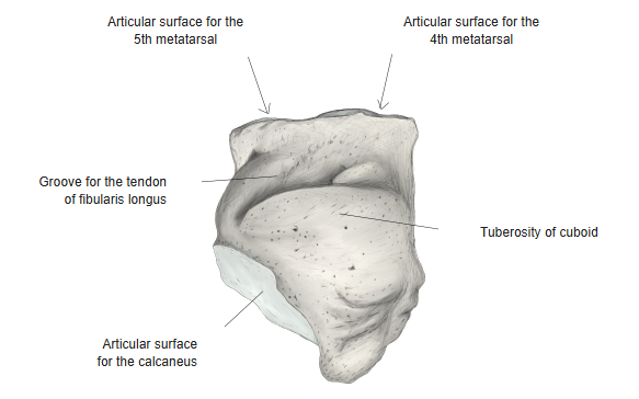
- From the lateral surface to the plantar surface runs the groove for the tendon of fibularis longus; the ridge just behind the groove is the tuberosity of the cuboid.
- Its posterior surface projects backwards at its plantar-medial corner as the calcaneal process, which supports the calcaneus.
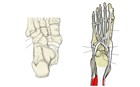
Groove for fibularis longus and tuberosity of the cuboid
- Bones of the lower limb (one side)
- Pelvic girdle
- Hip bone ← ilium, ischium and pubis fused
- Free lower limb
- Thigh and knee
- Femur
- Patella
- Leg
- Tibia ← big-toe side
- Fibula ← little-toe side
- Foot
- Tarsal bones (7)
- Metatarsal bones (5)
- Phalanges (14)
- Thigh and knee
- Pelvic girdle

Illustrations
Right foot (dorsal) 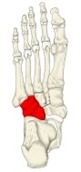
Right foot (plantar) 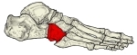
Right foot (lateral) 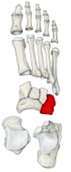
Bones of the foot (separated) 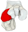
Tarsal bones (anterior) 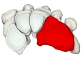
Distal tarsals and metatarsals (posterior) 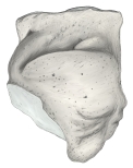
Right cuboid (plantar surface) 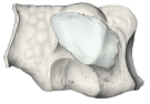
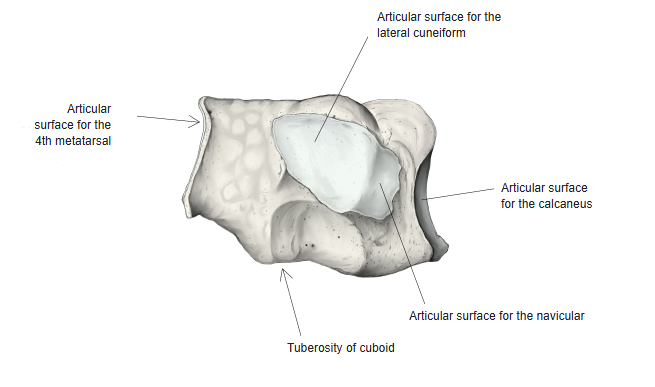
Right cuboid (medial surface) 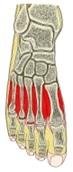
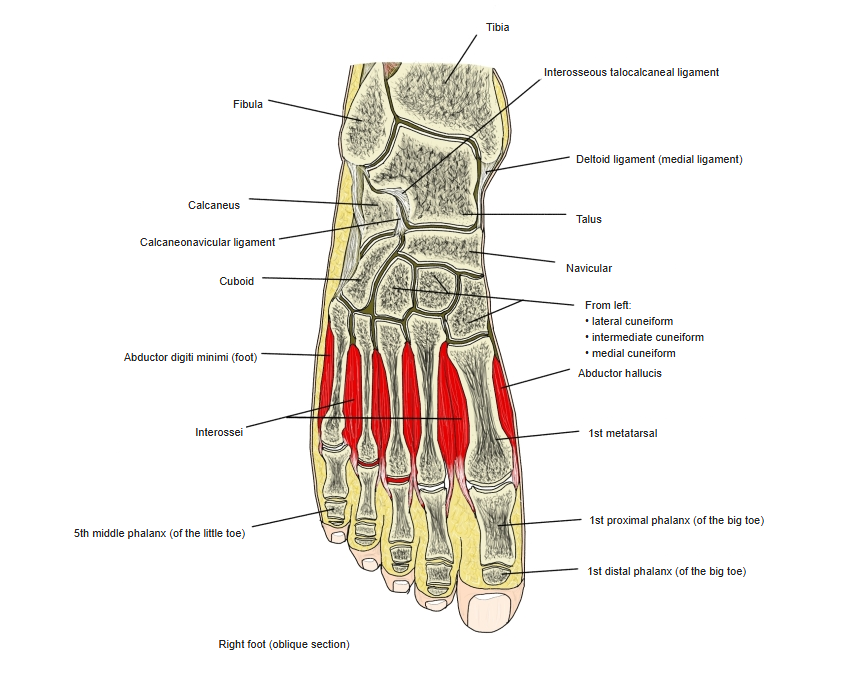
Right foot (oblique section)
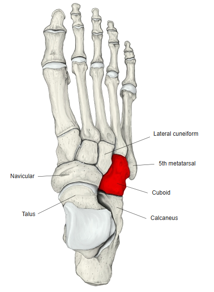
Articulations
| # | Bone | Joint |
|---|---|---|
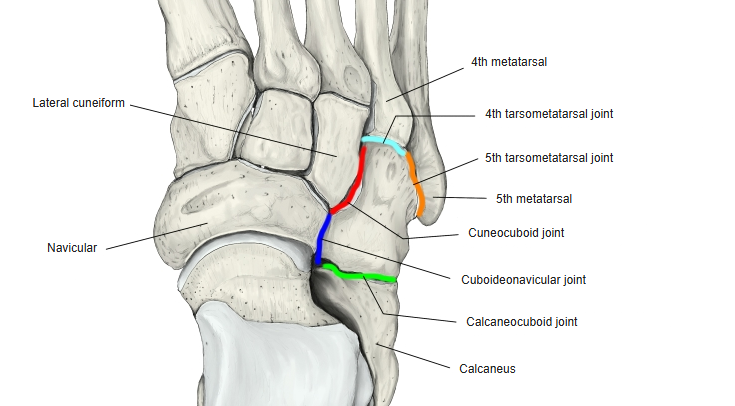
| 1 | 4th metatarsal | Tarsometatarsal joint (4th and 5th) |
| 2 | 5th metatarsal | |
| 3 | Lateral cuneiform | Cuneocuboid joint |
| 4 | Navicular | Cuboideonavicular joint |
| 5 | Calcaneus | Calcaneocuboid joint |
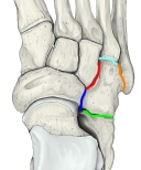
Joints of the cuboid
Ligaments
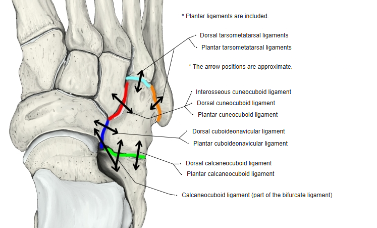
The following ligaments attach to the cuboid. The list is intended to be comprehensive but may omit minor ligaments.
| # | Ligament | Connects |
|---|---|---|
| 1 | Dorsal tarsometatarsal ligaments | Cuboid – 4th and 5th metatarsals |
| 2 | Plantar tarsometatarsal ligaments | Cuboid – 4th and 5th metatarsals |
| 3 | Dorsal cuneocuboid ligament | Cuboid – lateral cuneiform |
| 4 | Plantar cuneocuboid ligament | Cuboid – lateral cuneiform |
| 5 | Interosseous cuneocuboid ligament | Cuboid – lateral cuneiform |
| 6 | Dorsal cuboideonavicular ligament | Cuboid – navicular |
| 7 | Plantar cuboideonavicular ligament | Cuboid – navicular |
| 8 | Dorsal calcaneocuboid ligament | Cuboid – calcaneus |
| 9 | Plantar calcaneocuboid ligament | Cuboid – calcaneus |
| 10 | Calcaneocuboid ligament (part of the bifurcate ligament) | Cuboid – calcaneus |
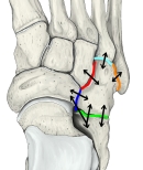
Ligaments of the cuboid
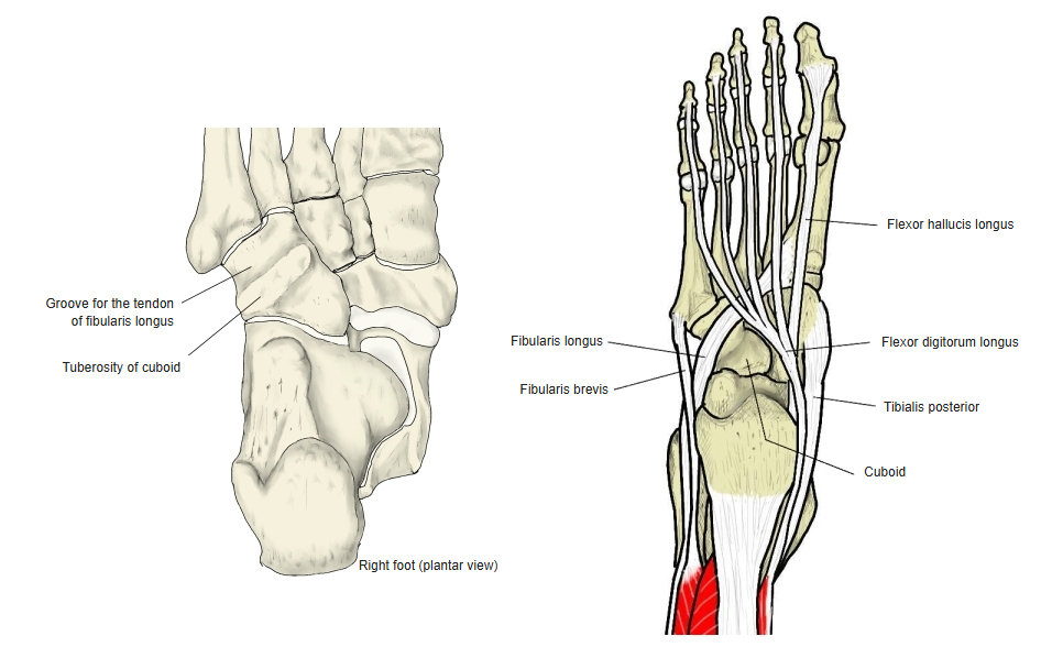
Muscle attachments
- Origin of: Flexor hallucis brevis, Adductor hallucis (oblique head)
- Insertion of: Tibialis posterior (a slip of its tendon)
Not a muscle, but the deep fibres of the long plantar ligament attach to the tuberosity of the cuboid.
References and sources
- Further reading: Cuboid bone, Wikipedia.