Latin: os coccygis · the tailbone
Overview
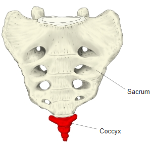
- The small bone at the lower end of the vertebral column, below the sacrum.
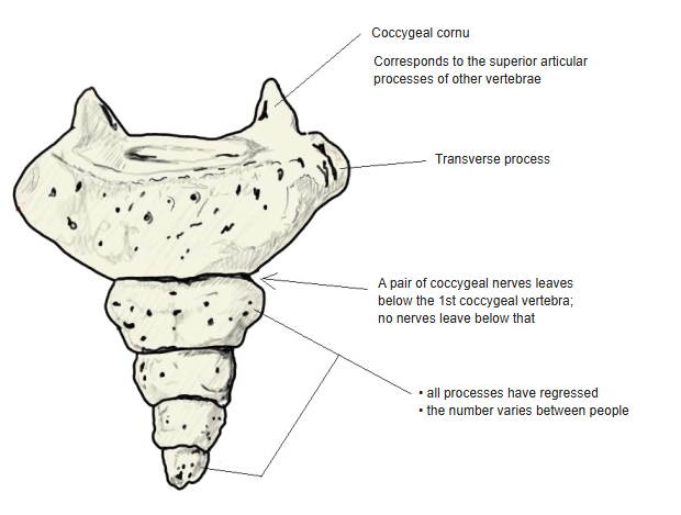
- The embryo has the primordia of about nine coccygeal vertebrae; the lower ones regress, and the upper three to five remain and form the coccyx, so their number varies between people.
- The segments are often described as fused, but in adults the coccyx may consist of up to five separate segments; two or three is the most common pattern.
- As a whole it is shaped like an inverted triangle.
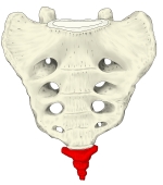
Coccyx and sacrum 
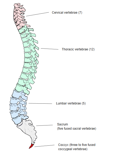
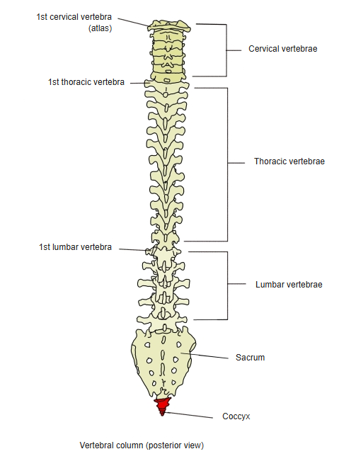
Coccyx in the vertebral column
- Bones of the trunk
- Vertebral column
- Cervical vertebrae (7)
- Thoracic vertebrae (12)
- Lumbar vertebrae (5)
- Sacrum (1)
- Coccyx (1)
- Thoracic cage
- Sternum (1)
- Ribs (24)
- Vertebral column
Illustrations

Vertebral column (posterior) 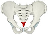
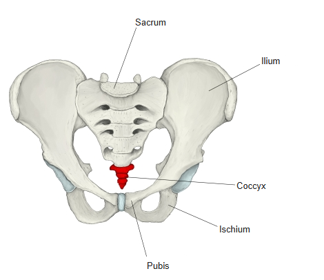
Pelvis (anterior) 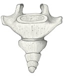
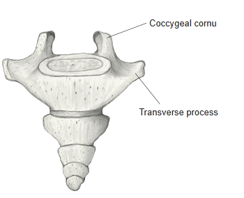
Coccyx (anterior) 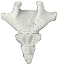
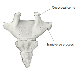
Coccyx (posterior) 
Pelvis (anterior oblique) 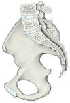
Pelvis (median section) 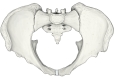
Pelvis (from above) 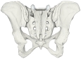
Pelvis (posterior) 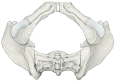
Pelvis (from below)
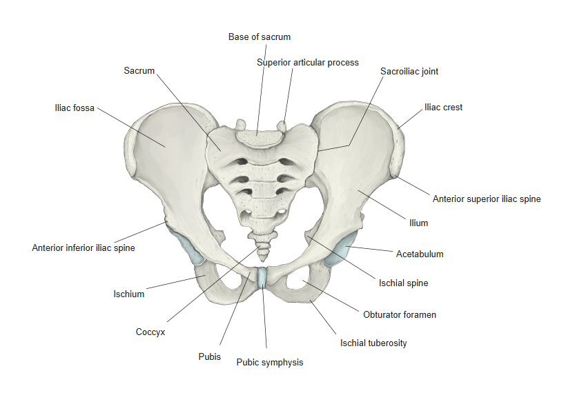
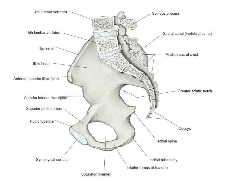
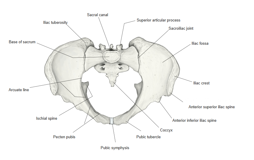
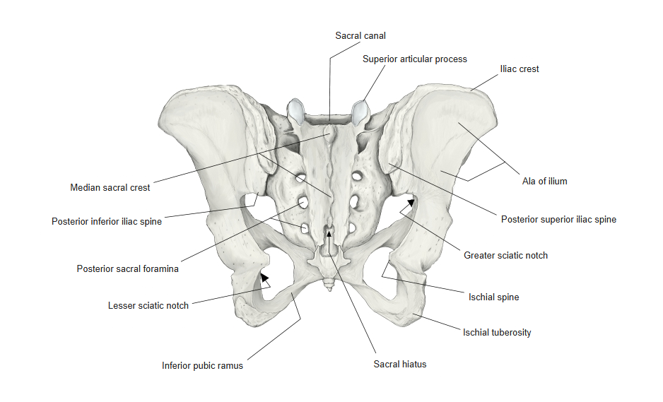
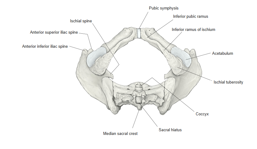
Articulations
| # | Bone | Joint |
|---|---|---|
| 1 | Sacrum (apex of sacrum) ⇔ 1st coccygeal vertebra | Sacrococcygeal joint |
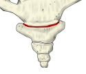
Sacrococcygeal joint
Ligaments
The following ligaments attach to the coccyx. The list is intended to be comprehensive but may omit minor ligaments.
| # | Ligament | Connects |
|---|---|---|
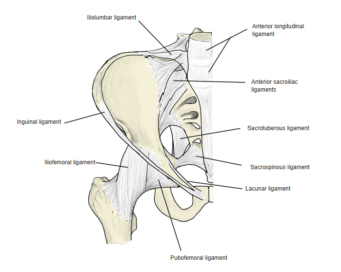
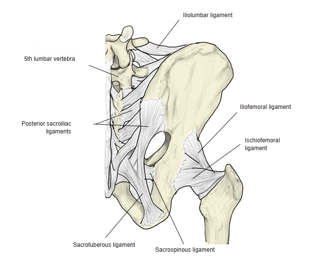
| 1 | Sacrotuberous ligament | Ischial tuberosity – coccyx, sacrum and posterior inferior iliac spine |
| 2 | Sacrospinous ligament | Ischial spine – lateral margins of the sacrum and coccyx |
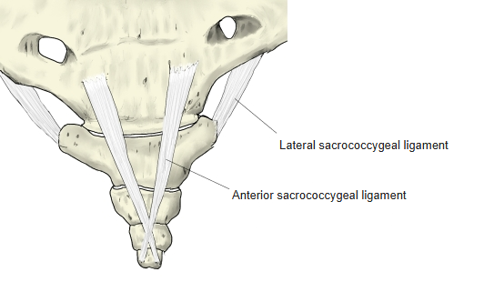
| 3 | Anterior sacrococcygeal ligament | Front of the sacrum and coccyx |
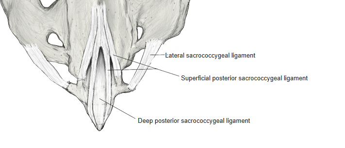
| 4 | Posterior sacrococcygeal ligament (superficial and deep) | Back of the sacrum and coccyx |
| 5 | Lateral sacrococcygeal ligament | Lateral edge of the apex of the sacrum – transverse process of the 1st coccygeal vertebra |
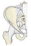
Ligaments of the pelvis (anterior) 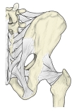
Ligaments of the pelvis (posterior) 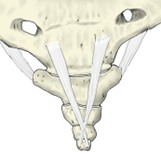
Anterior sacrococcygeal ligament 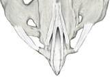
Posterior and lateral sacrococcygeal ligaments
Muscle attachments
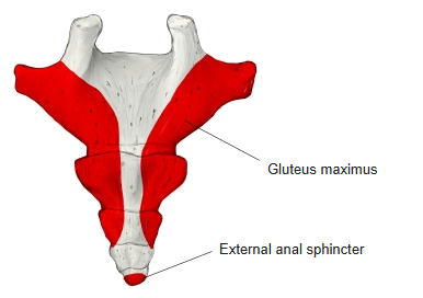
- Origin of: Gluteus maximus, External anal sphincter
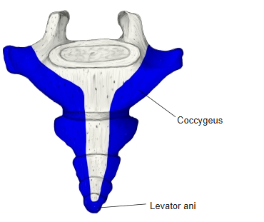
- Insertion of: Coccygeus, Sacrococcygeus anterior (ventralis), Sacrococcygeus posterior (dorsalis), Levator ani
Gluteus maximus and coccygeus: lateral margin · external anal sphincter and levator ani: tip of the coccyx · sacrococcygeus anterior / posterior: front / back.
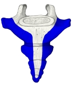
Muscle insertions (anterior) 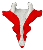
Muscle origins (posterior) 
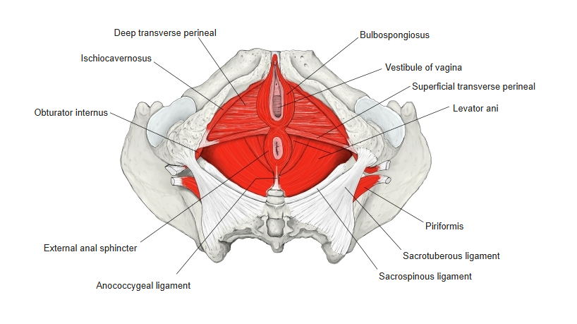
Muscles of the perineum and pelvic floor
Shape
- Upper part (1st coccygeal vertebra) – keeps some features of a vertebra: two upward projections, the coccygeal cornua (corresponding to superior articular processes), and short transverse processes at their bases.
- Lower segments – the processes have regressed.
- The 1st coccygeal nerve leaves below the 1st coccygeal vertebra; no nerves leave below that.
- If the coccyx is not fused with the sacrum, the two meet at the sacrococcygeal joint through an intervertebral disc.
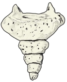
Shape of the coccyx
References and sources
- Further reading: Coccyx, Wikipedia.