Latin: mandibula
Overview
- The lower jaw: the largest and strongest bone of the face and the only movable bone of the skull (apart from the ear ossicles).
- It has a horseshoe-shaped body carrying the lower teeth and two vertical rami that rise from its back ends; body and ramus meet at the angle of the mandible.
- Each ramus ends above in two processes – the coronoid process in front and the condylar process behind, separated by the mandibular notch. The head of the condylar process forms the temporomandibular joint with the temporal bone.
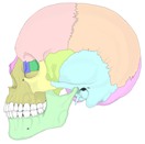
Bones of the skull (lateral) 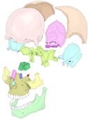
Bones of the skull (separated)
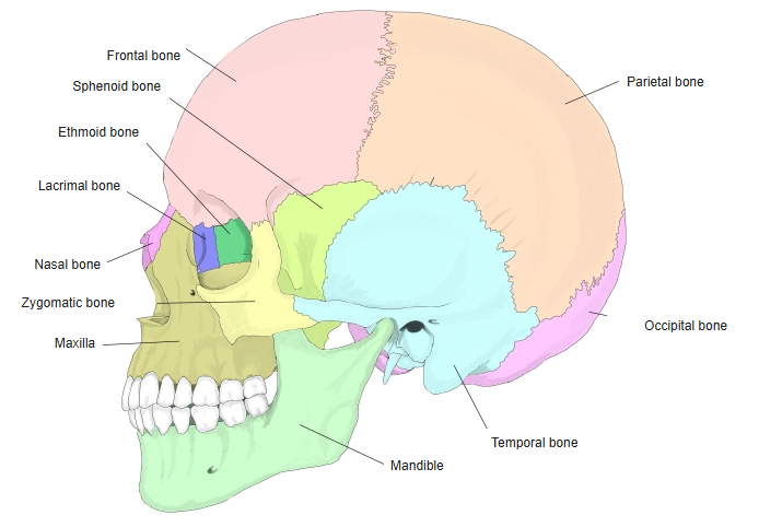
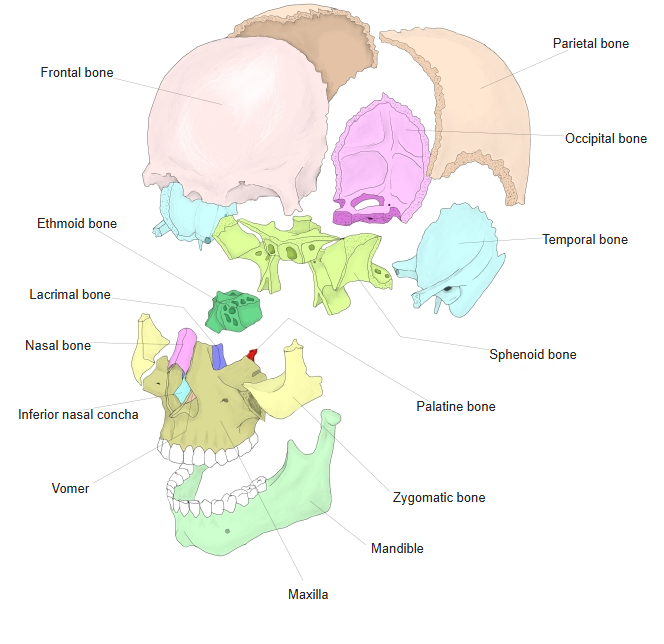
- Bones of the skull
- Neurocranium (cranial bones)
- Frontal bone
- Parietal bone (2)
- Occipital bone
- Temporal bone (2)
- Sphenoid bone
- Ethmoid bone
- Viscerocranium (facial bones)
- Nasal bone (2)
- Vomer
- Lacrimal bone (2)
- Inferior nasal concha (2)
- Maxilla (2)
- Zygomatic bone (2)
- Palatine bone (2)
- Mandible
- Hyoid bone
- Neurocranium (cranial bones)
Illustrations
Articulations
| # | Bone | Joint |
|---|---|---|
| 1 | Temporal bones | Temporomandibular joints (synovial joints) |
Ligaments
The following ligaments attach to the mandible. The list is intended to be comprehensive but may omit minor ligaments.
| # | Ligament | Connects |
|---|---|---|
| 1 | Lateral ligament (temporomandibular joint) | Zygomatic arch and articular tubercle – neck of mandible |
| 2 | Sphenomandibular ligament | Spine of sphenoid – lingula of mandible |
| 3 | Stylomandibular ligament | Styloid process – angle and posterior border of the ramus |
Muscle attachments
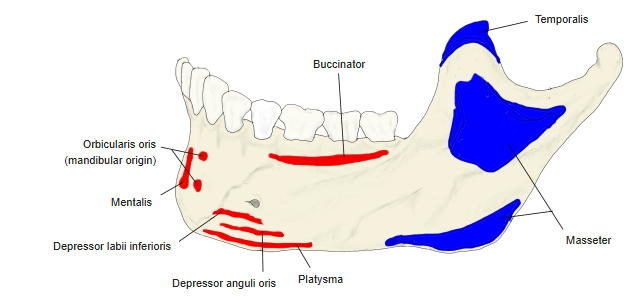
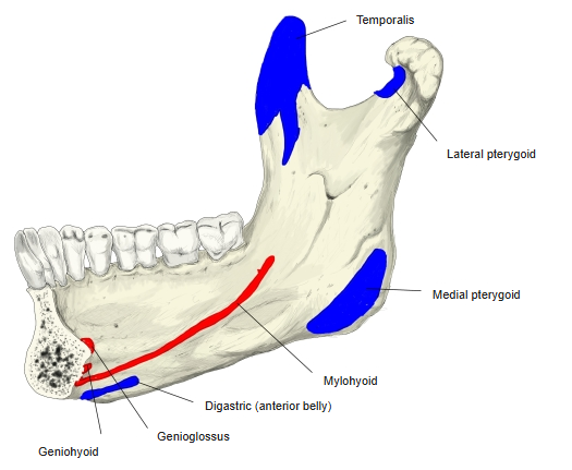
- Origin of: Buccinator, Mentalis, Depressor labii inferioris, Depressor anguli oris, Orbicularis oris (in part), Genioglossus, Geniohyoid, Mylohyoid, Digastric (anterior belly), Superior pharyngeal constrictor (in part)
- Insertion of: Temporalis, Masseter, Medial pterygoid, Lateral pterygoid, Platysma
In the figures, origins are shown in red and insertions in blue. The four muscles of mastication all insert on the mandible.
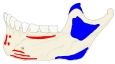
Muscle attachments (lateral) 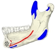
Muscle attachments (medial)
Body
- Mental protuberance and mental tubercles – the prominence of the chin in the midline.
- Mental foramen – below the second premolar; the mental nerve and vessels emerge here.
- Oblique line – runs up from the mental tubercle to the anterior border of the ramus.
- Alveolar part – the upper part, with the sockets of the lower teeth.
- Mental spines (genial tubercles) – on the inner surface in the midline, for genioglossus and geniohyoid.
- Mylohyoid line – a ridge on the inner surface for the mylohyoid; the sublingual fossa lies above it in front and the submandibular fossa below it behind.
- Digastric fossa – beside the midline on the lower border, for the anterior belly of digastric.
Ramus
- Coronoid process – a thin triangular plate in front, for the insertion of temporalis.
- Condylar process – the head of the mandible on a neck, with the pterygoid fovea on the front of the neck for the lateral pterygoid.
- Mandibular foramen – in the middle of the inner surface, guarded by the lingula; it leads into the mandibular canal for the inferior alveolar nerve and vessels. The mylohyoid groove runs down from it.
- Masseteric tuberosity and pterygoid tuberosity – roughened areas at the outer and inner angle for masseter and medial pterygoid.
References and sources
- Further reading: Mandible, Wikipedia.