Condylar process (Latin: processus condylaris mandibulae)
Also called the condyloid process
· It is thicker than the coronoid process.
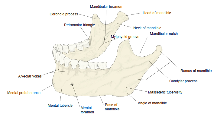
- Body of mandible : the flat, curved front part in which the roots of the lower teeth are set. Features: mental foramen, mental protuberance, mylohyoid line, alveolar part, etc.
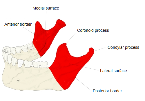
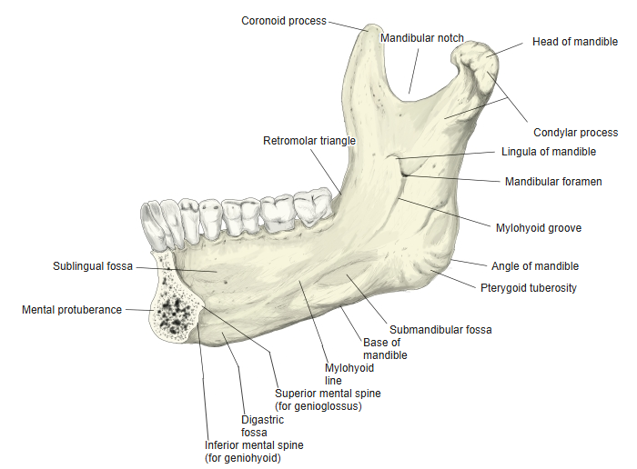
- Ramus of mandible : the paired back part, rising upwards and backwards on each side. Features: coronoid process, condylar process, mandibular notch, mandibular foramen, mylohyoid groove, etc.
Common to both parts: the angle of mandible and the mandibular canal.
- Head of mandible : the expanded upper end of the condylar process.
- Articular surface : oval; through the articular disc it articulates with the mandibular fossa of the temporal bone → temporomandibular joint
- Neck of mandible : the narrowed part below the head.
- Pterygoid fovea : a small pit on the front of the neck; insertion of the lateral pterygoid.
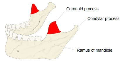
The mandibular notch lies between the condylar process and the coronoid process in front of it.
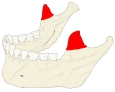 Coronoid and condylar processes
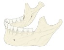 Mandible (oblique view)
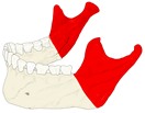 Ramus of mandible
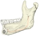 Right half of mandible (medial view)
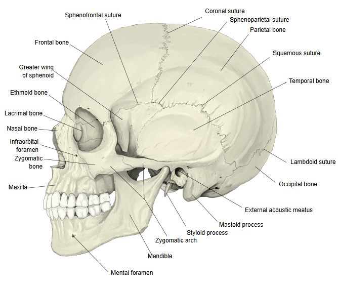
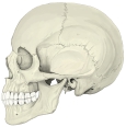 Skull (lateral view)
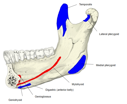
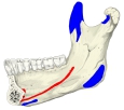 Mandible: muscle attachments (medial surface)
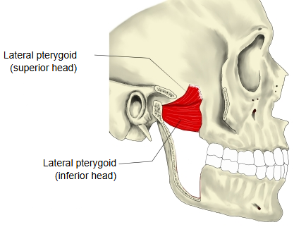
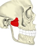 Lateral pterygoid
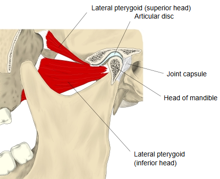
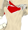 Temporomandibular joint (sagittal section)
In more detail:
The upper end of the ramus divides into two processes: the thin coronoid process in front and the thicker condylar process behind, separated by the mandibular notch. The condylar process has two parts: the expanded head (the mandibular condyle) at the top and the narrower neck that supports it.
Head of mandible: oval and elongated from side to side; its articular surface is convex from front to back and from side to side. Its long axis points medially and slightly backwards, so that the axes of the two heads meet near the front edge of the foramen magnum. The articular surface is covered by fibrocartilage (dense fibrous tissue) rather than hyaline cartilage. Through the articular disc the head articulates with the mandibular fossa and the articular tubercle of the temporal bone; when the mouth opens wide, the head and the disc glide forwards onto the tubercle. A small tubercle on its lateral end gives attachment to the lateral ligament of the joint.
Neck of mandible: the narrowed part below the head, flattened from front to back and strengthened by ridges that run down from the head. On its front is a small depression, the pterygoid fovea, where the lateral pterygoid inserts (mainly its inferior head; some fibres of the superior head reach the articular disc and capsule).
Fractures of the condylar neck are common after a blow to the chin.
Reference websites
[Wikipedia]
· Condyloid process, Wikipedia.
· Temporomandibular joint, Wikipedia.
[Websites with illustrations]
· Condyloid process, lateral view (Wikimedia Commons)

· Condyloid process, close-up, superior view (Wikimedia Commons)

· Articulation of the mandible, lateral aspect (Gray's Anatomy, fig. 309) (Wikimedia Commons)

· The temporomandibular joint (TeachMeAnatomy)
Where it is found
Related terms
- Ramus of mandible
- Coronoid process
- Temporomandibular joint
- Angle of mandible
- Mandibular foramen
- Mandible
References and sources
- Further reading: Condyloid process, Wikipedia.
- Further reading: Temporomandibular joint, Wikipedia.
- Federative International Programme for Anatomical Terminology (FIPAT). Terminologia Anatomica, 2nd ed. (TA2), 2019.