Trochlea of humerus (Latin: trochlea humeri)
· "Condyle of humerus = trochlea of humerus + capitulum of humerus" (some sources also count the radial fossa, coronoid fossa and so on as part of the condyle).
· It articulates with the trochlear notch of the ulna ⇒ humeroulnar joint (one part of the elbow joint).
On the lower surface of the distal end, between the medial and lateral epicondyles, is the condyle of the humerus, the articular head that meets the radius and the ulna. A low ridge divides it into a medial and a lateral part. The medial part is the trochlea of the humerus, which articulates with the ulna (the trochlear notch).
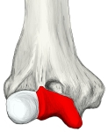 Trochlea (anterior)
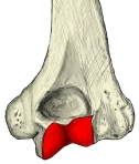 Trochlea (posterior)
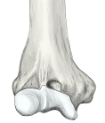 Lower end (anterior)
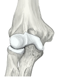 Right elbow (anterior)
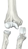 Elbow bones, separated
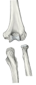 Elbow bones (posterior)
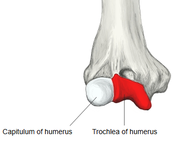
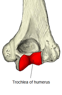
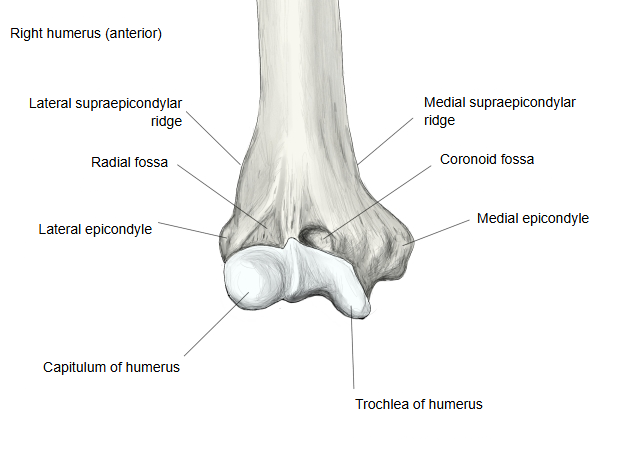
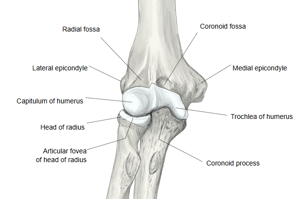
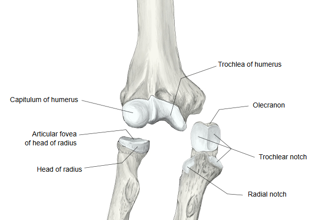
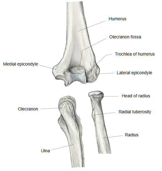
The trochlea is shaped like a pulley: a deep central groove lies between two raised borders, and the medial border projects further down than the lateral. It covers the front, the lower surface and the back of the condyle, with the capitulum on its lateral side and the medial epicondyle on its medial side. Above it lie the coronoid fossa in front and the olecranon fossa behind, which take the coronoid process and the olecranon in full flexion and extension; occasionally the bone between them is perforated (the supratrochlear foramen). Its groove runs spirally, which is one reason the extended forearm is angled slightly outwards from the arm (the carrying angle, usually larger in women). With the trochlear notch it forms the humeroulnar joint, a hinge joint and the main part of the elbow joint. Its centre of ossification appears late, at about 8-9 years of age.
Where it is found
Related terms
- Condyle of humerus
- Capitulum of humerus
- Coronoid fossa
- Olecranon fossa
- Trochlear notch
- Humeroulnar joint
- Distal epiphysis of humerus
References and sources
- Further reading: Trochlea of humerus, Wikipedia.
- Further reading: Carrying angle, Wikipedia.
- Federative International Programme for Anatomical Terminology (FIPAT). Terminologia Anatomica, 2nd ed. (TA2), 2019.