Pubic symphysis (Latin: symphysis pubica)
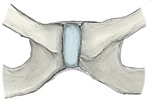 Pubic symphysis (anterior)
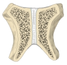 Pubic symphysis (coronal section)
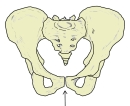 Pelvis (anterior)
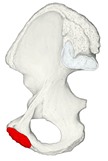 Right hip bone (medial)
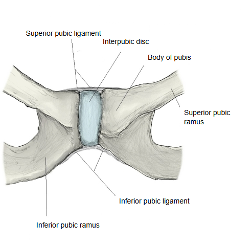
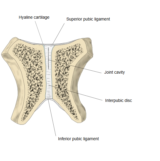
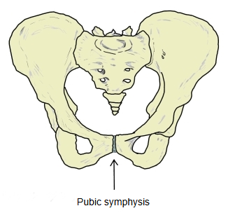
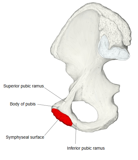
In the midline at the front of the pelvis, the two symphyseal surfaces, each covered by thin hyaline cartilage, come close together and are joined by the fibrocartilaginous interpubic disc, whose structure resembles the anulus fibrosus of an intervertebral disc. The joint lies in front of the urinary bladder and above the external genitalia. In pregnancy it is loosened, which helps the baby's head pass through the birth canal; in guinea pigs and some other animals it can be loosened experimentally by giving female sex hormones.
The pubic symphysis is a midline secondary cartilaginous joint (a symphysis) of the bony pelvis that unites the bodies of the two pubic bones (pubis (symphyseal surface) ⇔ interpubic disc ⇔ pubis (symphyseal surface)). A wedge-shaped fibrocartilaginous interpubic disc lies between the two thin layers of hyaline cartilage that line the oval symphyseal surfaces; the disc often contains a small slit-like cavity. The width of the joint space changes with age:
· it is widest in early childhood and narrows steadily through adult life.
The disc is thicker in women, which allows a little more movement of the pelvic bones and a slightly larger pelvic diameter for childbirth. The joint is reinforced by ligaments:
· Superior pubic ligament: runs along the top of the joint, between the pubic crests as far as the pubic tubercles.
· Inferior pubic ligament (arcuate or subpubic ligament): arches under the joint between the inferior pubic rami and forms the top of the pubic arch.
· Anterior pubic ligament: fibres in front of the joint that blend with the periosteum at the sides and with the interpubic disc.
· Posterior pubic ligament: thinner fibres behind the joint that blend with the periosteum of both pubic bodies.
(The anterior and posterior pubic ligaments are not named in every textbook; some sources treat these fibres as periosteum and the edge of the disc. Sources differ.)
Muscles and tendons
The adductor longus, adductor brevis and rectus abdominis attach close to the joint, with fibres blending into the anterior pubic ligament and the interpubic disc.
The aponeurosis of the external oblique also reinforces the joint in front.
Relations
In front:
the proximal parts of the shafts of the penis or clitoris.
Below:
the urethra;
the deep dorsal vein of the penis or of the clitoris.
Behind:
the urinary bladder, separated from the joint by the retropubic fat;
in men, the prostate and the prostatic venous plexus.
Movement: normally very small, about 2 mm of shift and 1° of rotation. In pregnancy hormones loosen the joint, letting the pelvis widen slightly for childbirth. Blood supply
Mainly branches of the obturator artery and the inferior epigastric artery.
Nerve supply
Branches of the pudendal nerve, and/or the genitofemoral, iliohypogastric and ilioinguinal nerves.
Variants
Congenital widening of the pubic symphysis (rare).
Imaging (CT and MRI)
In adolescents the joint can look V-shaped on axial images because of secondary ossification centres at the front; in adults the joint surfaces are normally parallel.
Related conditions
Pelvic instability after childbirth.
Widening of the pubic symphysis.
Open-book fracture of the pelvis.
Osteitis pubis: inflammation of the joint and nearby bone, a cause of groin pain in athletes.
Fusion (bridging) of the pubic symphysis.
Reference websites
[Wikipedia]
· Pubic symphysis, Wikipedia.
[Websites with illustrations]
· Symphysis pubis exposed by a coronal section (Gray's Anatomy) (Wikimedia Commons)

Where it is found
Related terms
References and sources
- Further reading: Pubic symphysis, Wikipedia.
- Federative International Programme for Anatomical Terminology (FIPAT). Terminologia Anatomica, 2nd ed. (TA2), 2019.