Medial intercondylar tubercle (Latin: tuberculum intercondylare mediale)
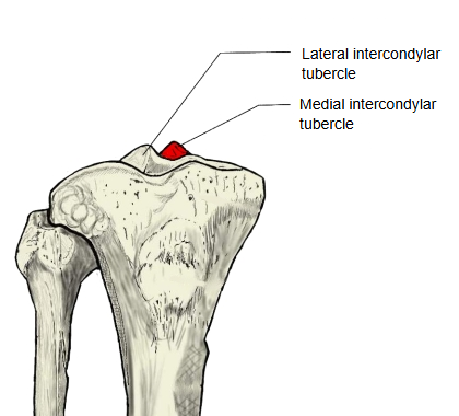
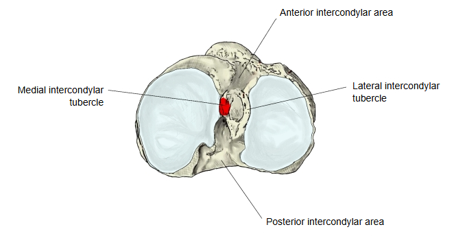
The intercondylar eminence, near the middle of the upper surface of the tibia, bears a medial and a lateral tubercle; the medial one is the medial intercondylar tubercle.
It is a small peak at the inner edge of the medial part of the superior articular surface, whose cartilage extends up onto its side. Its partner is the lateral intercondylar tubercle, separated from it by a small notch; the anterior and posterior intercondylar areas lie in front of and behind the pair. It projects into the intercondylar fossa of the femur.
- Medial intercondylar tubercle : the tubercle on the medial side of the intercondylar eminence.
- Lateral intercondylar tubercle : the tubercle on the lateral side of the intercondylar eminence.
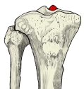 Right tibia and fibula (anterior)
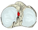 Right tibia (superior view)
Reference websites
[Wikipedia]
· Intercondylar area, Wikipedia.
· Tibia, Wikipedia.
[Websites with illustrations]
· Upper surface of the right tibia (Gray's Anatomy) (Wikimedia Commons)

· Bones of the right leg, anterior surface (Gray's Anatomy) (Wikimedia Commons)

· The tibia (TeachMeAnatomy)
Where it is found
Related terms
- Intercondylar eminence
- Lateral intercondylar tubercle
- Anterior intercondylar area
- Posterior intercondylar area
- Medial condyle of tibia
- Superior articular surface of tibia
References and sources
- Further reading: Intercondylar area, Wikipedia.
- Further reading: Tibia, Wikipedia.
- Federative International Programme for Anatomical Terminology (FIPAT). Terminologia Anatomica, 2nd ed. (TA2), 2019.