Intercondylar eminence (Latin: eminentia intercondylaris)
Also called the tibial spine
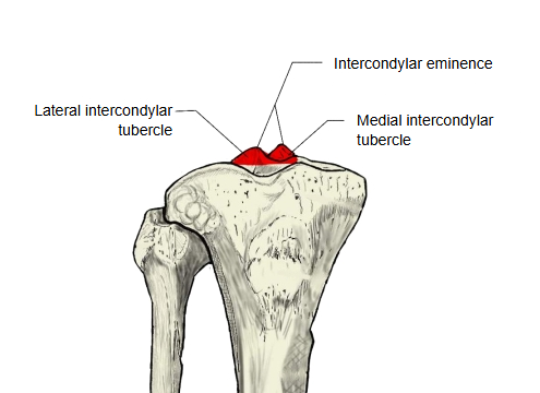
The raised area in the middle of the upper end of the tibia (the end that meets the femur at the knee), between the two superior articular surfaces. Its top rises into two small peaks: the medial one is the medial intercondylar tubercle and the lateral one the lateral intercondylar tubercle.
eminence
- Medial intercondylar tubercle : the peak on the medial side of the intercondylar eminence.
- Lateral intercondylar tubercle : the peak on the lateral side of the intercondylar eminence.
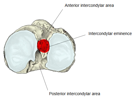
It lies between the medial and lateral condyles, a little nearer the back of the bone than the front. The articular surfaces of the condyles extend up onto the sides of the two tubercles.
In front of it is the anterior intercondylar area and behind it the posterior intercondylar area. The anterior and posterior cruciate ligaments and the horns of the medial and lateral menisci attach to these rough areas rather than to the eminence itself.
When the knee is straightened it fits into the intercondylar fossa of the femur. Note: a fracture of the intercondylar eminence (tibial spine avulsion), seen mostly in children, pulls off the bony attachment of the anterior cruciate ligament.
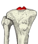 Right tibia and fibula (anterior)
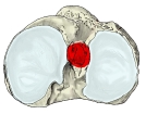 Right tibia (superior view)
Reference websites
[Wikipedia]
· Intercondylar area, Wikipedia.
· Tibia, Wikipedia.
[Websites with illustrations]
· Upper surface of the right tibia (Gray's Anatomy, fig. 257) (Wikimedia Commons)

· Tibia: anatomy and clinical notes (Kenhub)
· The tibia: proximal, shaft and distal anatomy (TeachMeAnatomy)
Where it is found
Related terms
- Medial intercondylar tubercle
- Lateral intercondylar tubercle
- Anterior intercondylar area
- Posterior intercondylar area
- Superior articular surface of tibia
- Proximal epiphysis of tibia
- Intercondylar fossa
References and sources
- Further reading: Intercondylar area, Wikipedia.
- Further reading: Tibia, Wikipedia.
- Federative International Programme for Anatomical Terminology (FIPAT). Terminologia Anatomica, 2nd ed. (TA2), 2019.