Superior articular surface of tibia (Latin: facies articularis superior tibiae)
Also called the tibial plateau
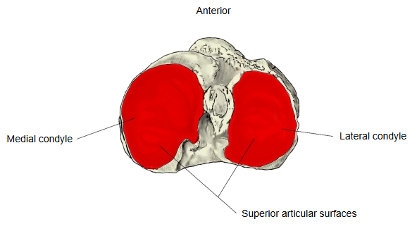
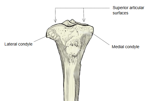
The slightly hollowed, cartilage-covered surfaces on top of the medial condyle and the lateral condyle of the tibia. They face the medial and lateral condyles of the femur and form the knee joint with them.
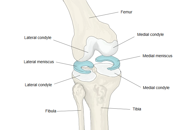
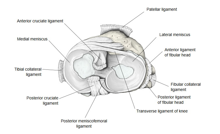
On each superior articular surface rests one of the menisci, the medial meniscus and the lateral meniscus, which deepen the shallow surfaces for the rounded femoral condyles.
surfaces of
the tibia
- Superior articular surface : the joint surfaces on top of the medial and lateral condyles → knee joint.
- Fibular articular facet : on the back and underside of the lateral condyle → tibiofibular joint.
- Inferior articular surface : on the underside of the lower end → talocrural (ankle) joint.
- Articular surface of medial malleolus : on the lateral side of the medial malleolus, continuous with the inferior articular surface → talocrural (ankle) joint.
The two surfaces differ. The medial surface is larger and oval, longer from front to back, and slightly concave. The lateral surface is smaller and more nearly circular; it is concave from side to side but flat or slightly convex from front to back. Both extend up onto the sides of the intercondylar tubercles.
They are separated by the intercondylar eminence and by the rough intercondylar areas in front of and behind it. The fibrous capsule of the knee joint attaches close to their outer margins.
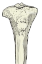 Upper end of the right tibia (anterior view)
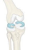 Right knee joint with the menisci (anterior view)
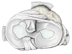 Medial meniscus and lateral meniscus (superior view)
Reference websites
[Wikipedia]
· Tibia, Wikipedia.
· Meniscus (anatomy), Wikipedia.
[Websites with illustrations]
· Upper surface of the right tibia (Gray's Anatomy, fig. 257) (Wikimedia Commons)

· Tibia: anatomy and clinical notes (Kenhub)
· The tibia: proximal, shaft and distal anatomy (TeachMeAnatomy)
Where it is found
Related terms
- Medial condyle of tibia
- Lateral condyle of tibia
- Intercondylar eminence
- Proximal epiphysis of tibia
- Fibular articular facet
- Inferior articular surface of tibia
- Knee joint
References and sources
- Further reading: Tibia, Wikipedia.
- Further reading: Meniscus (anatomy), Wikipedia.
- Federative International Programme for Anatomical Terminology (FIPAT). Terminologia Anatomica, 2nd ed. (TA2), 2019.