Posterior intercondylar area (Latin: area intercondylaris posterior)
The area on the upper surface of the tibia behind the intercondylar eminence, near the middle of the bone; it is where the posterior cruciate ligament (PCL) attaches.
· It lies between the back parts of the two superior articular surfaces and slopes down towards the back of the bone.
· From front to back it receives the posterior horn of the lateral meniscus (just behind the lateral intercondylar tubercle), the posterior horn of the medial meniscus, and the posterior cruciate ligament.
· The posterior cruciate ligament attaches at its back edge and extends a little onto the back of the tibia. It runs upwards, forwards and medially to the inner side of the medial condyle of the femur.
· In front of the eminence is the matching anterior intercondylar area.
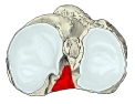 Right tibia (superior view)
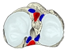 Right tibia (superior view): ligament and meniscus attachments
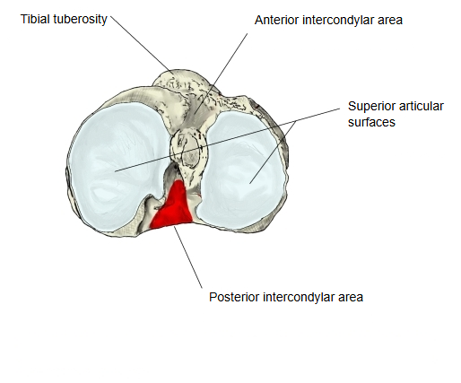
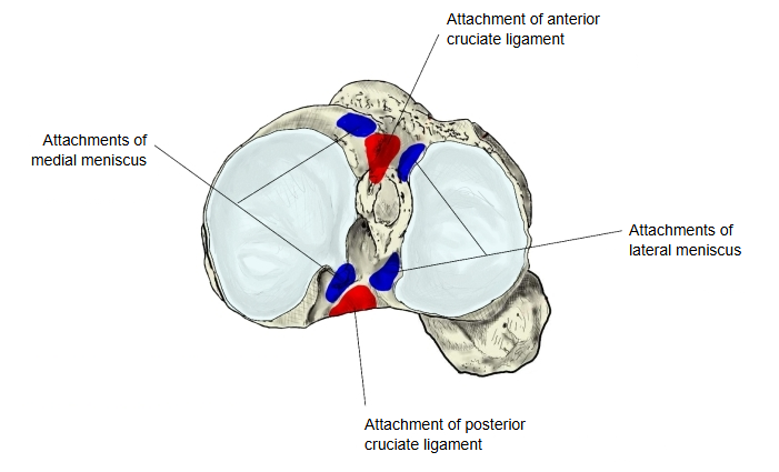
Attached structures
| Ligament | 1 | Posterior cruciate ligament | ligamentum cruciatum posterius |
The extent of the red area and the attachment sites in the figures are approximate.
Reference websites
[Wikipedia]
· Intercondylar area, Wikipedia.
· Posterior cruciate ligament, Wikipedia.
[Websites with illustrations]
· Upper surface of the right tibia (Gray's Anatomy, fig. 257) (Wikimedia Commons)

· The tibia (TeachMeAnatomy)
Where it is found
Related terms
- Anterior intercondylar area
- Intercondylar eminence
- Medial intercondylar tubercle
- Lateral intercondylar tubercle
- Superior articular surface of tibia
- Proximal epiphysis of tibia
References and sources
- Further reading: Intercondylar area, Wikipedia.
- Further reading: Posterior cruciate ligament, Wikipedia.
- Federative International Programme for Anatomical Terminology (FIPAT). Terminologia Anatomica, 2nd ed. (TA2), 2019.