Lateral malleolus (Latin: malleolus lateralis)
· It is the bony bump on the outer side of the ankle, often called the 'outer ankle bone', and is easy to feel under the skin.
· The corresponding projection on the inner side (the part of the lower end of the tibia that projects downwards on the medial side) is the medial malleolus (the 'inner ankle bone').
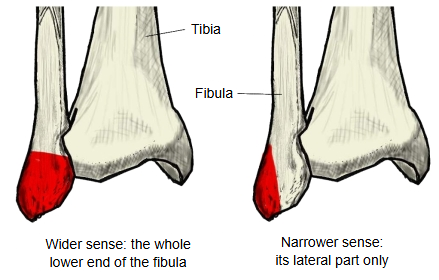 Two uses of the term
 Right ankle (anterior)
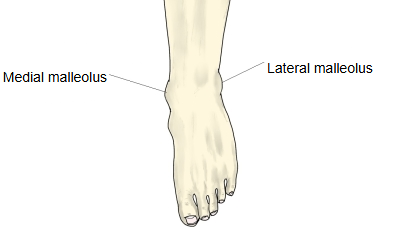
The lower end of the fibula, the lateral malleolus, is slightly enlarged and roughly four-sided, with a pointed tip. The upper part of its medial side is joined to the tibia. Below this lies a triangular, cartilage-covered surface, the articular surface of the lateral malleolus, which meets the talus at the talocrural (ankle) joint. Behind it is the malleolar fossa, from which the posterior talofibular ligament arises; the calcaneofibular ligament arises from the front of the tip. On its back is a groove for the tendons of fibularis longus and fibularis brevis (the malleolar groove).
The lower end of the fibula is thickened and projects downwards as the lateral malleolus. It reaches lower than the medial malleolus and lies further back, and its tip points downwards and backwards rather than straight down. The name usually covers the whole projecting lower end of the fibula (distal epiphysis); a few texts restrict it to its lateral part.
Attached structures
The list may differ a little depending on whether the lateral malleolus is taken as the whole enlarged lower end or only its lateral part; the ligaments attached to the enlarged lower end of the fibula are listed below.
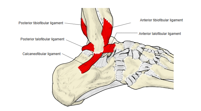
| Ligaments | 1 | Anterior tibiofibular ligament | fibula (lower end, probably the lateral malleolus) ⇔ tibia (lower end) |
| 2 | Posterior tibiofibular ligament | fibula (lower end, probably the lateral malleolus) ⇔ tibia (lower end) | |
| 3 | Anterior talofibular ligament | fibula (lower end: lateral malleolus) ⇔ talus (neck of talus) | |
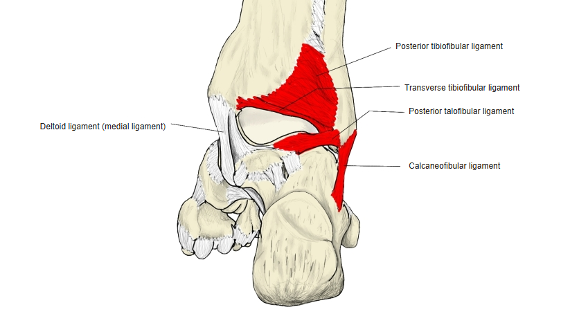
| 4 | Posterior talofibular ligament | fibula (lower end: malleolar fossa) ⇔ talus (lateral tubercle) | |
| 5 | Transverse tibiofibular ligament | fibula (lower end, probably the lateral malleolus) ⇔ tibia (lower end) | |
| 6 | Calcaneofibular ligament | fibula (lower end: lateral malleolus, near its tip) ⇔ calcaneus (lateral surface) |
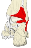 Right ankle ligaments (posterior)
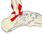 Right ankle ligaments (lateral)
Clinical notes
· The anterior talofibular ligament is the ligament most often torn in an inversion ankle sprain.
· Fractures of the lateral malleolus are among the most common ankle fractures.
Reference websites
[Wikipedia]
· Malleolus, Wikipedia.
· Fibula, Wikipedia.
· Ankle, Wikipedia.
[Websites with illustrations]
· Bones of the right leg, anterior surface (Gray's Anatomy) (Wikimedia Commons)

· Ligaments of the foot, lateral aspect (Gray's Anatomy) (Wikimedia Commons)

· Fibula: anatomy and bone landmarks (Kenhub)
Where it is found
Related terms
- Distal epiphysis of fibula
- Articular surface of lateral malleolus
- Malleolar fossa
- Malleolar groove of fibula
- Medial malleolus
- Talocrural joint
- Lateral malleolar facet