Medial malleolus (Latin: malleolus medialis)
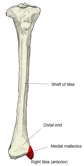
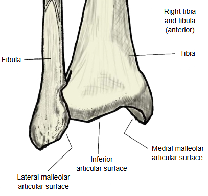
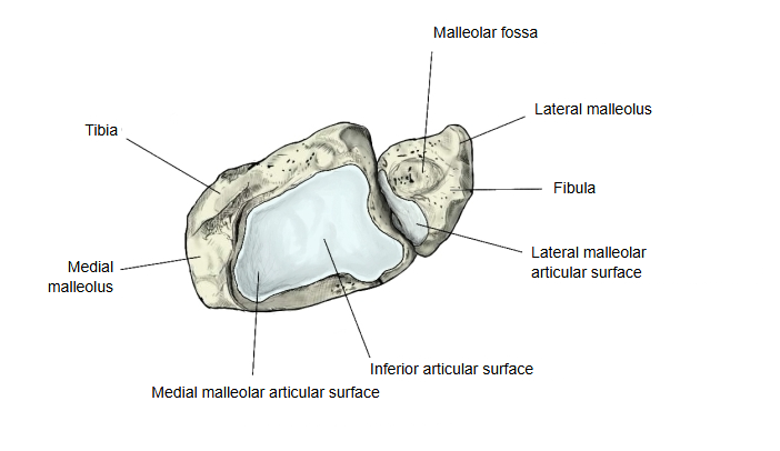
The part of the lower end of the tibia that projects downwards on its medial side. It is the inner ankle bone, easily felt under the skin. Its medial surface is rough, while its lateral surface, the articular surface of the medial malleolus, articulates with the talus at the talocrural (ankle) joint.
It is shorter than the lateral malleolus of the fibula and lies a little higher and further forward. The malleolar groove on its back carries the tendon of tibialis posterior. Note: the medial malleolus is often broken in ankle injuries, sometimes together with the lateral malleolus (bimalleolar fracture).
Attachments
| Ligament | 1 | Deltoid ligament | medial ligament of the ankle; attached to the tip and borders of the medial malleolus |
Reference websites
[Wikipedia]
· Malleolus, Wikipedia.
· Tibia, Wikipedia.
[Websites with illustrations]
· Lower end of the tibia, posterior view (photograph) (Wikimedia Commons)

· Bones of the right leg, anterior surface (Gray's Anatomy, fig. 258) (Wikimedia Commons)

· Tibia: anatomy and clinical notes (Kenhub)
Where it is found
Related terms
- Articular surface of medial malleolus
- Malleolar groove of tibia
- Inferior articular surface of tibia
- Distal epiphysis of tibia
- Lateral malleolus
- Talocrural joint