Medial process of calcaneal tuberosity (Latin: processus medialis tuberis calcanei)
- Attachments
- Reference websites
- (Wikipedia, websites with illustrations)
- Where it is found
- Related terms
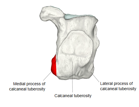
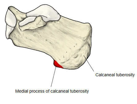
A process on the medial side of the underside of the calcaneal tuberosity of the calcaneus; beside it, on the other side, is the smaller lateral process of the calcaneal tuberosity. It is the larger of the two processes. Anatomy websites describe it as follows.
It is a broad projection on the plantar (lower) surface of the calcaneal tuberosity. It is broader than the lateral process and reaches lower (further towards the sole), so it takes most of the weight of the heel in standing. Muscles of the sole arise from it, and the flexor retinaculum and the plantar aponeurosis are attached to it.
· Heel pain from plantar fasciitis, and bony heel spurs, are usually found at the medial process.
Attachments
| 1 | Muscles | 1 | Abductor hallucis |
| 2 | Flexor digitorum brevis | ||
| 3 | Abductor digiti minimi (part) | ||
| 2 | Others | 1 | Flexor retinaculum (to its medial side) |
| 2 | Plantar aponeurosis (central part) |
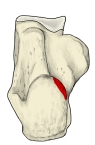 Right calcaneus (plantar)
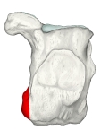 Right calcaneus (posterior)
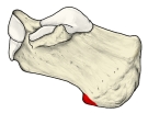 Right calcaneus (medial)
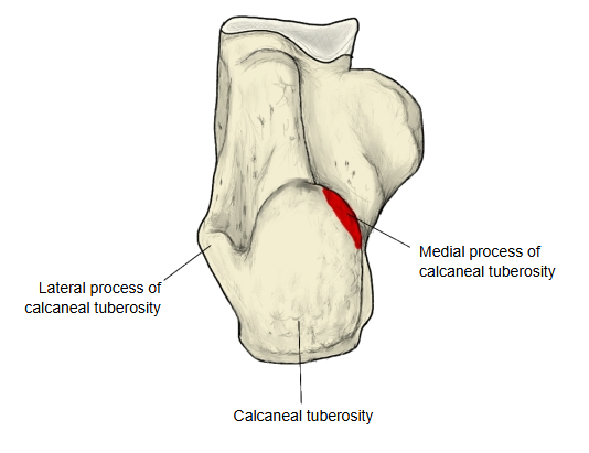
Reference websites
[Wikipedia]
· Calcaneus, Wikipedia.
· Plantar fascia, Wikipedia.
[Websites with illustrations]
· Left calcaneus, medial surface (Gray's Anatomy, fig. 267) (Wikimedia Commons)

· Left calcaneus, inferior surface (Gray's Anatomy, fig. 265) (Wikimedia Commons)

Where it is found
Related terms
- Calcaneal tuberosity
- Lateral process of calcaneal tuberosity
- Body of calcaneus
- Calcaneal tubercle
- Sustentaculum tali
References and sources
- Further reading: Calcaneus, Wikipedia.
- Further reading: Plantar fascia, Wikipedia.
- Federative International Programme for Anatomical Terminology (FIPAT). Terminologia Anatomica, 2nd ed. (TA2), 2019.