Jugular foramen (Latin: foramen jugulare)
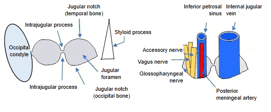
The foramen lies at the back end of the petro-occipital fissure, lateral to the hypoglossal canal and the occipital condyle and behind the carotid canal. The jugular notch of the petrous part of the temporal bone forms its front and lateral margin, that of the occipital bone its back and medial margin, and the two meet like two halves of a ring. The intrajugular processes are often joined by fibrous tissue or a bony bridge. On the outer skull base the foramen opens into the jugular fossa of the temporal bone, a deep hollow for the superior bulb of the internal jugular vein. The foramen is usually larger on the right, matching the larger right sigmoid sinus.
Photographs of many skulls show that the way the intrajugular processes of the two bones meet is not the same in everyone. The following cases are seen:
· the tips of the two processes lie almost on one line;
· the tips are clearly offset from each other;
· the processes are too small to be called processes;
· one (or both) of the processes is missing.
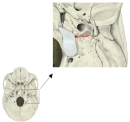 External cranial base (enlarged)
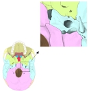 External cranial base, bones coloured
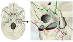 Left jugular foramen: jugular notches and intrajugular processes
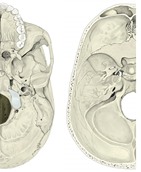 Internal and external cranial base
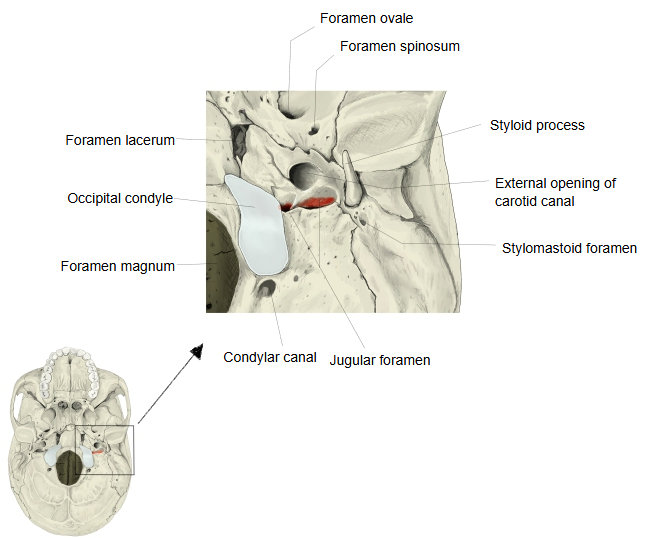
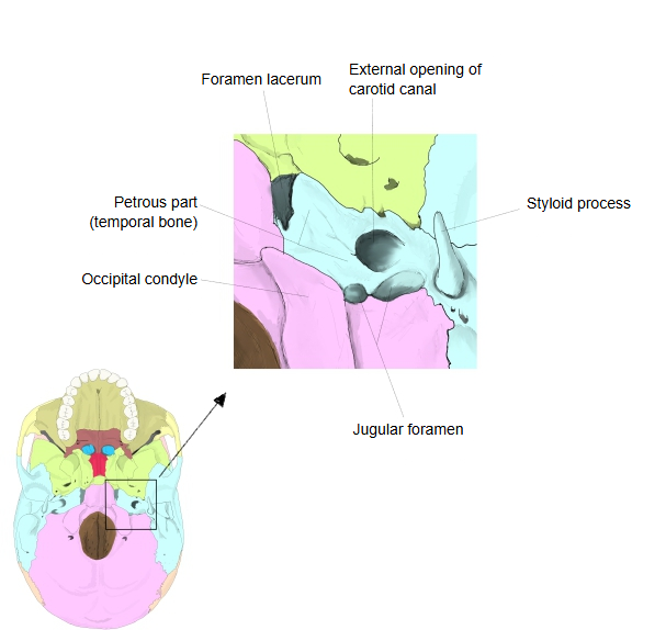
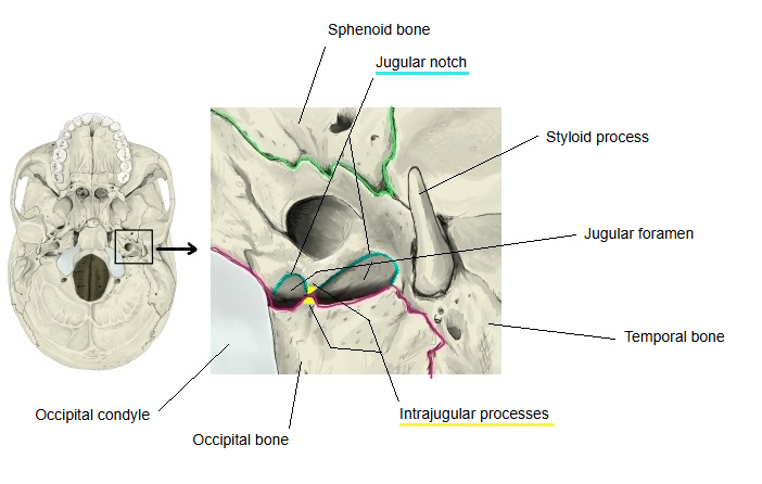
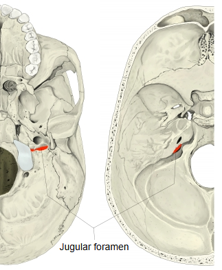
Structures passing through
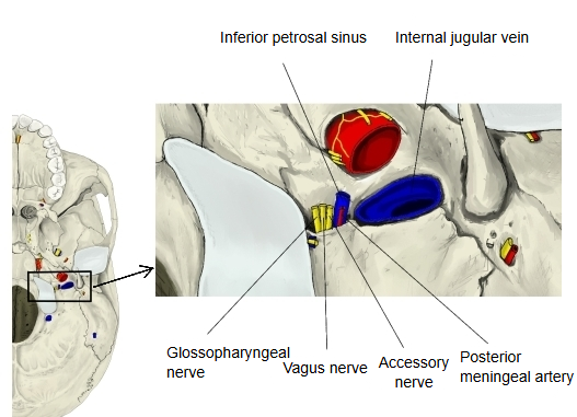
The following nerves and vessels pass through the jugular foramen.
| Name | Notes | |
| Nerves | Glossopharyngeal nerve | Cranial nerve IX (ninth of the cranial nerves) |
| Vagus nerve | Cranial nerve X | |
| Accessory nerve | Cranial nerve XI | |
| Veins | Internal jugular vein | Begins here as the continuation of the sigmoid sinus; passes through the larger lateral (posterolateral) part, separated off by the intrajugular processes. |
| Inferior petrosal sinus | Passes through the front (anteromedial) part. | |
| Arteries | Posterior meningeal artery | A branch of the ascending pharyngeal artery. |
| Occipital artery (meningeal branches) | May enter instead through the condylar canal, the hypoglossal canal or the mastoid foramen. |
Many texts describe three compartments: a front one (inferior petrosal sinus), a middle one (cranial nerves IX, X and XI) and a back one (sigmoid sinus / internal jugular vein and the small arteries); sources differ in the details. Tumours or fractures at the foramen can damage nerves IX, X and XI together (jugular foramen syndrome).
 Nerves and vessels passing through (external view)
 Nerves and vessels passing through (internal view)
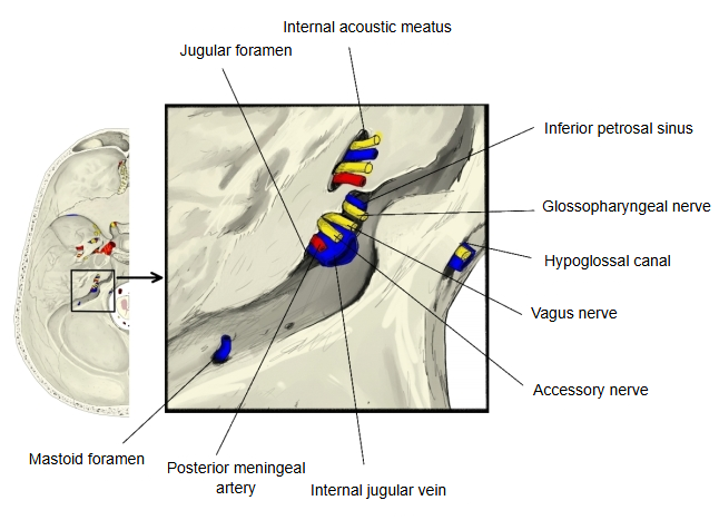
· Some sources also list emissary veins and the occipital artery among the structures that pass through the larger lateral part of the foramen.
Reference websites
[Wikipedia]
· Jugular foramen, Wikipedia.
· Jugular foramen syndrome, Wikipedia.
[Websites with illustrations]
· Jugular foramen (Kenhub)
· Base of the skull, external surface (Gray's Anatomy) (Wikimedia Commons)

· Base of the skull, internal surface (Gray's Anatomy) (Wikimedia Commons)

Where it is found
Related terms
- Hypoglossal canal
- Foramen magnum
- Occipital condyle
- Petrous part
- Carotid canal
- Occipital bone
- Temporal bone
References and sources
- Further reading: Jugular foramen, Wikipedia.
- Further reading: Jugular foramen syndrome, Wikipedia.
- Federative International Programme for Anatomical Terminology (FIPAT). Terminologia Anatomica, 2nd ed. (TA2), 2019.