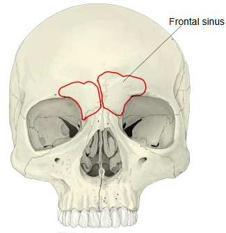
Frontal sinus (Latin: sinus frontalis)
It has the following features.
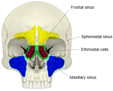
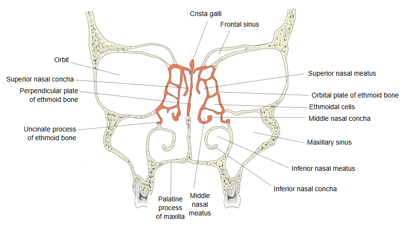
- Maxillary sinus : in the maxilla; the largest of the paranasal sinuses.
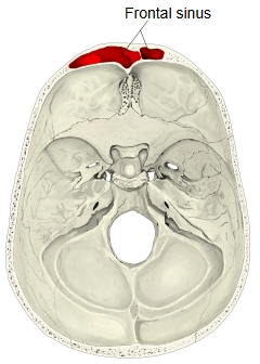
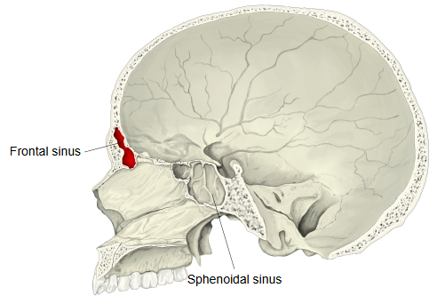
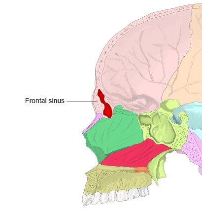
- Frontal sinus : in the frontal bone.
- Ethmoidal sinus : in the ethmoid bone, divided into many small spaces (ethmoidal cells).
- Sphenoidal sinus : in the sphenoid bone, behind and deep to the ethmoidal sinus.
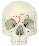 Skull (anterior)
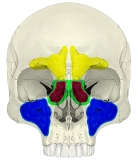 Paranasal sinuses (anterior)
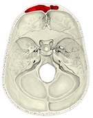 Cranial base (internal surface)
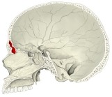 Skull (sagittal section)
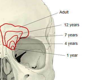
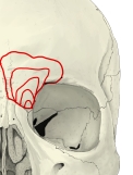 Growth of the frontal sinus
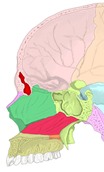 Skull (sagittal section, bones coloured)
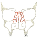 Nasal cavity (coronal section)
Other
An older German anatomy textbook (Rauber-Kopsch) describes the neighbouring bones as follows.
Among the bones that border the orbital part of the frontal bone, the right and left ethmoidal labyrinths come first: the roof of each labyrinth is formed by the medial part of the orbital part. Next is the lacrimal bone, set between the orbital plate of the ethmoid and the frontal process of the maxilla. Along the junction with the orbital plate lie the ethmoidal foveolae, which form the tops of the ethmoidal cells (ethmoidal sinus). Here there are two notches that the orbital plate closes into canals: the larger front one becomes the orbitocranial canal (anterior ethmoidal canal) and the smaller back one the orbitoethmoidal canal (posterior ethmoidal canal). Sometimes these canals are formed by the frontal bone alone or, more rarely, by the orbital plate alone.
Where it is found
Related terms
- Superciliary arch
- Glabella
- Frontal squama
- Maxillary sinus
- Sphenoidal sinus
- Ethmoidal air cells
- Frontal bone
References and sources
- Further reading: Frontal sinus, Wikipedia.
- Further reading: Paranasal sinuses, Wikipedia.
- Federative International Programme for Anatomical Terminology (FIPAT). Terminologia Anatomica, 2nd ed. (TA2), 2019.