Parietal layer of glomerular capsule
Also called the parietal epithelium
Overview
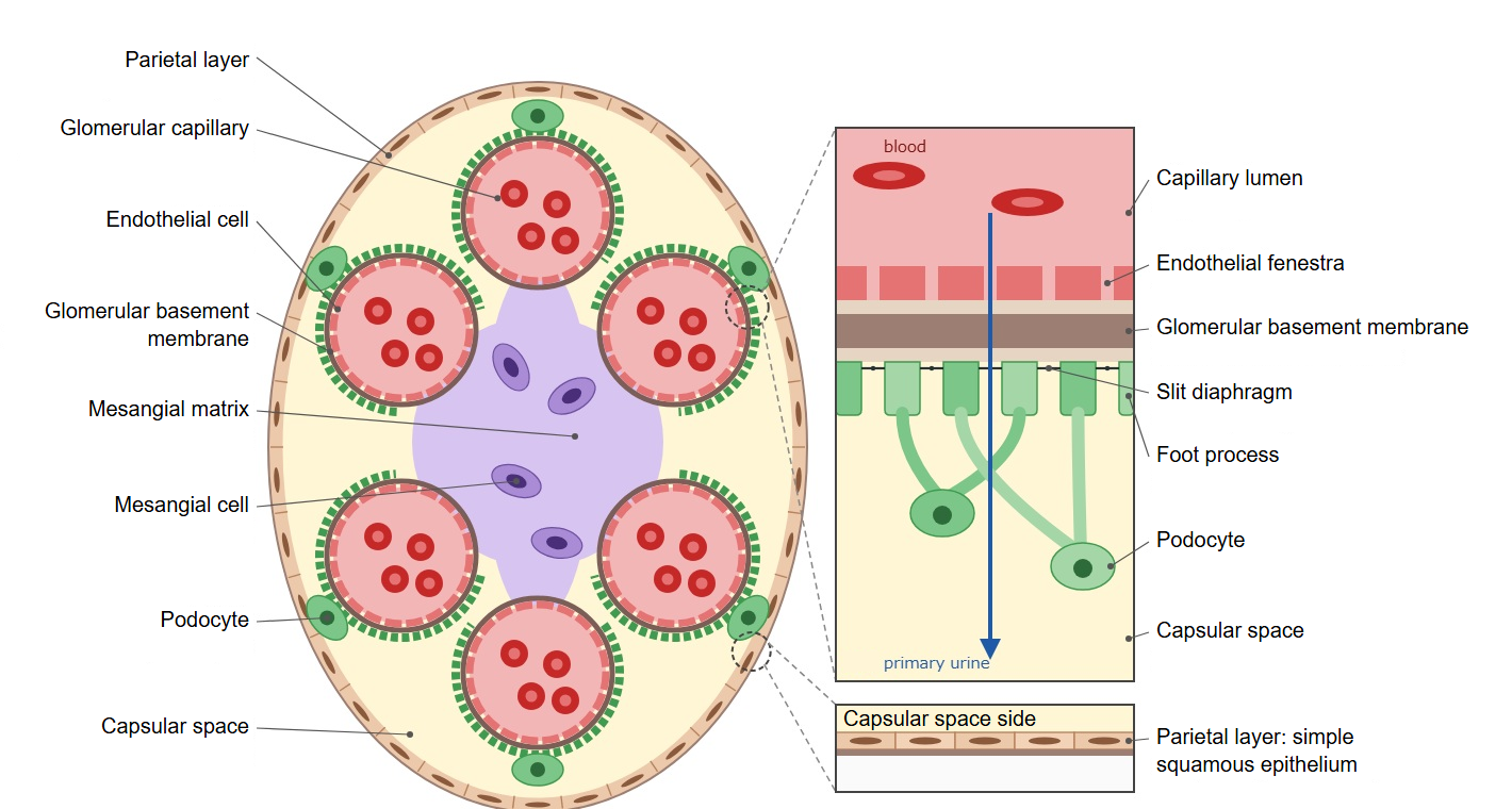
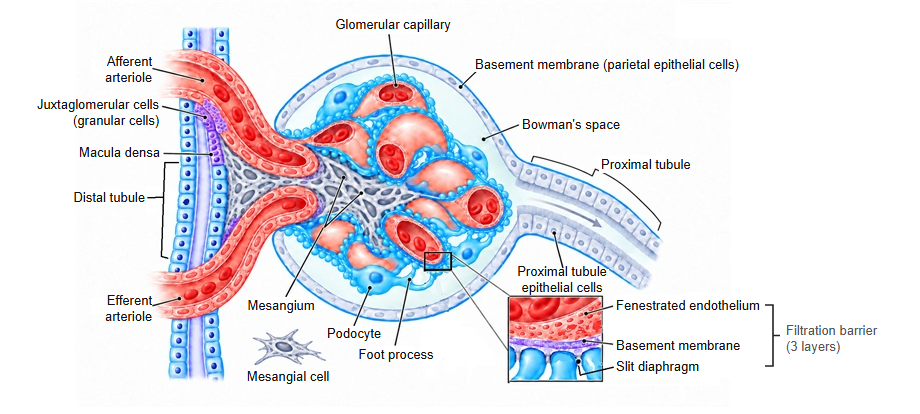
· The parietal layer is the outer layer of the glomerular capsule (Bowman's capsule), a thin wall of simple squamous epithelium. It is also called the parietal epithelium.
· It forms the outer wall of the cup that encloses the glomerulus. On its inner side it faces the visceral layer across the capsular space. The visceral layer is made of podocytes, but the parietal layer is made of ordinary flat cells.
· At the vascular pole it turns back and becomes continuous with the visceral layer; at the opposite urinary pole it continues into the epithelium of the proximal tubule.
· The parietal layer takes no part in filtering the blood. It is the wall that encloses the capsular space and guides the filtrate into the proximal tubule.
· In rapidly progressive glomerulonephritis, the parietal epithelial cells multiply and form crescent-shaped masses (crescents) in the capsular space.
Reference websites
[Wikipedia]
· Bowman's capsule, Wikipedia.
· Renal corpuscle, Wikipedia.
[Websites with illustrations]
· Renal corpuscle (Digital Histology)
· Renal corpuscle (detail) (Digital Histology)
· Renal corpuscle (diagram) (Wikimedia Commons)

Related terms
- Glomerular capillary
- Glomerular basement membrane
- Podocyte
- Mesangium
- Visceral layer of glomerular capsule
- Capsular space
- Vascular pole
- Urinary pole
- Proximal tubule
- Kidney
References and sources
- Further reading: Bowman's capsule, Wikipedia.
- Further reading: Renal corpuscle, Wikipedia.
- Federative International Programme for Anatomical Terminology (FIPAT). Terminologia Anatomica, 2nd ed. (TA2), 2019.