Connecting tubule (Latin: tubulus reuniens)
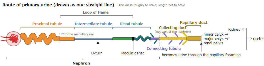
Also called the junctional tubule or arcuate renal tubule (CNT)
Overview
· It is also called the junctional tubule and is abbreviated CNT.
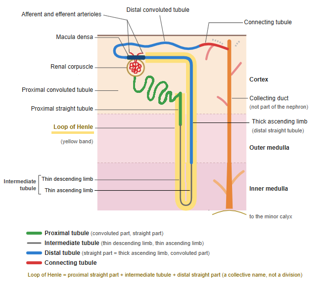
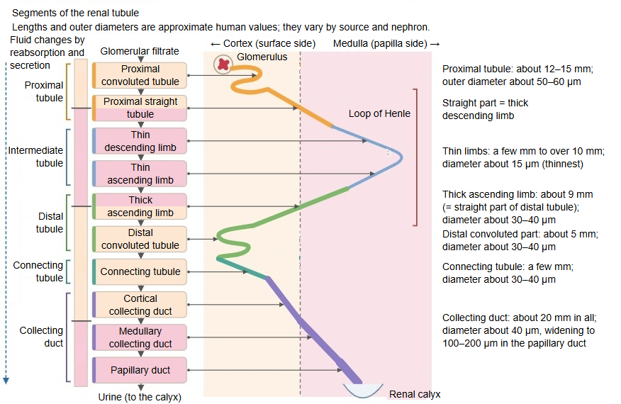
· It lies in the cortical labyrinth of the renal cortex. The connecting tubules of several nephrons join to form a collecting duct. In juxtamedullary nephrons the connecting tubules may first rise in arches and unite, forming arcades (arched collecting tubules).
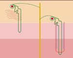
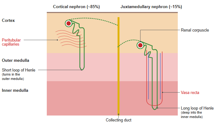
- Cortical nephron : one by one, each drains directly into a collecting duct
- Juxtamedullary nephron : several join to form an arcade (arched collecting tubule), which drains into a collecting duct
cortex
- Cortical labyrinth : renal corpuscles, proximal convoluted tubules, distal convoluted tubules, connecting tubules, juxtaglomerular apparatus, interlobular arteries and veins, afferent and efferent arterioles, capillariesabout 70–75%
- Medullary rays : proximal straight tubules, thick ascending limbs of the loop of Henle, collecting ducts, capillariesabout 25–30%
The percentages are approximate and differ between sources.
· It is thought to develop from the same metanephric tissue as the nephron, unlike the collecting duct, which grows from the ureteric bud. Sources therefore differ: some count it as part of the nephron, others as part of the collecting duct system.
· Its epithelium is intermediate between those of the distal convoluted tubule and the collecting duct: connecting tubule cells (similar to the principal cells of the collecting duct) are mixed with intercalated cells. It takes part in sodium reabsorption and potassium secretion under aldosterone, and in water reabsorption under antidiuretic hormone.
· Older descriptions joined the distal convoluted tubule and this segment as the intermediate (interposed) part and called this segment its second part; they regarded it as the transition to the collecting part of the tubule. This older "intermediate part" is not the modern intermediate tubule.
 Cortical and juxtamedullary nephrons
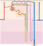 Nephron and blood vessels
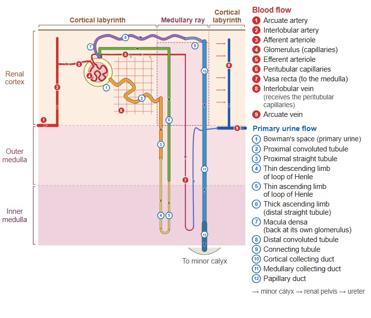
Reference websites
[Wikipedia]
· Collecting duct system, Wikipedia.
[Websites with illustrations]
· Cells of the nephron (Wikimedia Commons)

· Physiology of the nephron (Wikimedia Commons)

· Gray's Anatomy, figure 1128 (Wikimedia Commons)

· Histology of the tubules (kidneypathology.com)
Related terms
- Distal convoluted tubule
- Cortical collecting duct
- Collecting duct
- Cortical labyrinth
- Principal cell
- Intercalated cell
- Nephron
References and sources
- Further reading: Collecting duct system, Wikipedia.
- Federative International Programme for Anatomical Terminology (FIPAT). Terminologia Anatomica, 2nd ed. (TA2), 2019.