Intercalated cell (Latin: epitheliocytus intercalatus)
Also called the dark cell
Overview
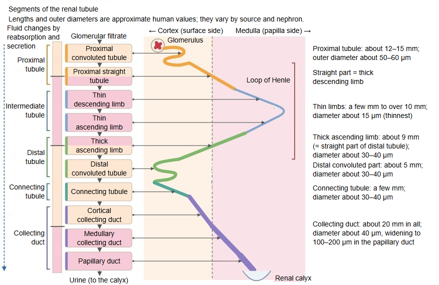
· Intercalated cells are cells of the epithelium of the connecting tubule and collecting duct of the kidney, scattered singly among the principal cells. Because they look dark they are also called dark cells. They secrete hydrogen ions (acid) or bicarbonate ions (base) and so help to control the pH (acid-base balance) of the body fluids.
· They are fewer than the principal cells and become rarer deeper in the renal medulla; in the papillary ducts there are few or none among the pale principal cells.
· Their cytoplasm is packed with mitochondria and stains darkly, and the luminal surface carries folds and many microvilli. The cells often bulge into the lumen.
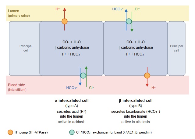
· Alpha (type A) intercalated cells make hydrogen and bicarbonate ions inside the cell with carbonic anhydrase. They secrete the hydrogen ions into the lumen (primary urine, which later becomes urine) through pumps in the luminal membrane (H⁺-ATPase and others) and return the bicarbonate to the blood through an exchanger in the basal membrane (the Cl⁻/HCO₃⁻ exchanger band 3). Their activity rises when the body becomes acidic (acidosis).
· Beta (type B) intercalated cells work in the opposite direction: they release bicarbonate into the lumen through pendrin, a Cl⁻/HCO₃⁻ exchanger in the luminal membrane, and are active when the body becomes alkaline (alkalosis). A third, non-alpha non-beta type also exists.
· If the alpha cells cannot secrete acid properly, distal (type 1) renal tubular acidosis results. The term has nothing to do with the intercalated duct of glands such as the pancreas.
Reference websites
[Wikipedia]
· Intercalated cell, Wikipedia.
[Websites with illustrations]
· Tubules and collecting system (collecting duct) (Digital Histology)
· Tubules and collecting system (collecting duct cells) (Digital Histology)
· Alpha intercalated cell (Wikimedia Commons)

· Tubular reabsorption (OpenStax Anatomy and Physiology 2e)
Related terms
- Principal cell
- Collecting duct
- Connecting tubule
- Cortical collecting duct
- Medullary collecting duct
- Papillary duct
- Kidney
References and sources
- Further reading: Intercalated cell, Wikipedia.
- Federative International Programme for Anatomical Terminology (FIPAT). Terminologia Anatomica, 2nd ed. (TA2), 2019.