Corneal limbus (Latin: limbus corneae)
Also called the corneoscleral junction
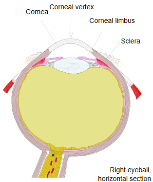
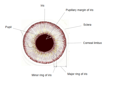
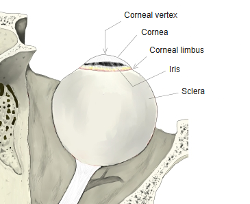
· The corneal limbus is the ring-shaped border zone where the transparent cornea passes into the opaque white sclera. The sclera overlaps the edge of the cornea slightly, so on the outside the border lies in a shallow groove (scleral sulcus). Deep to it lie the trabecular meshwork and the scleral venous sinus, through which aqueous humour drains, so it is an important landmark in eye surgery.
· The limbus is rich in blood vessels. Here the stratified squamous epithelium of the cornea becomes continuous with the epithelium of the conjunctiva.
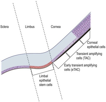
· The limbus holds the limbal stem cells in small radial fibrovascular ridges, the palisades of Vogt; they renew the corneal epithelium throughout life.
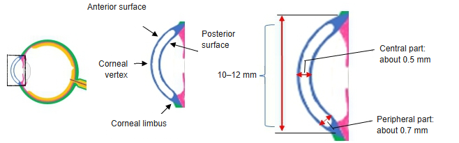
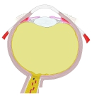 Eyeball (horizontal section)
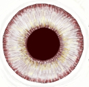 Iris and pupil (front view)
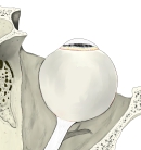 Right eyeball (superior view)
Reference websites
[Wikipedia]
· Corneal limbus, Wikipedia.
· Limbal stem cell, Wikipedia.
[Websites with illustrations]
· Eyeball with the limbus between cornea and sclera (diagram) (Wikimedia Commons)

· Limbal epithelial stem cells between sclera and cornea (diagram) (Entokey)
Related terms
References and sources
- Further reading: Corneal limbus, Wikipedia.
- Further reading: Limbal stem cell, Wikipedia.
- Federative International Programme for Anatomical Terminology (FIPAT). Terminologia Anatomica, 2nd ed. (TA2), 2019.