Latin: organum visus
Overview
- The organ of sight: it receives light, forms an image and sends the information to the brain.
- Made up of the eyeball, the optic nerve and the accessory structures that move, protect and moisten the eye.
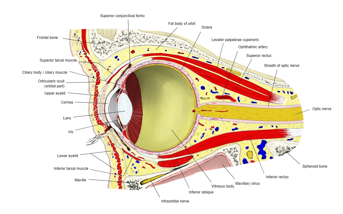
- Lies in the bony orbit, cushioned by orbital fat; the optic nerve leaves it through the optic canal.
- The optic nerve carries the signals to the brain; the visual pathway ends in the visual cortex of the occipital lobe.
Orbit, longitudinal section 
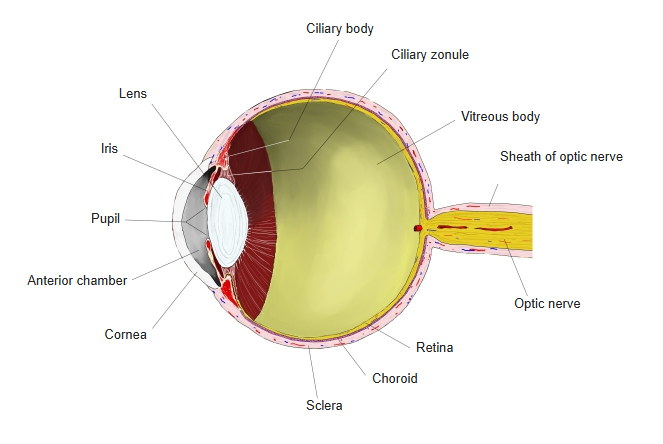
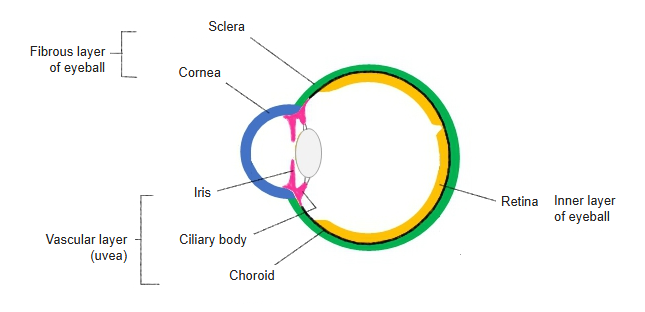
Eyeball, longitudinal section
Pages on the visual organ
- Accessory structures of eye
- Aqueous humor
- Chambers of eyeball
- Choroid
- Ciliary body
- Conjunctiva
- Cornea
- Eyeball
- Eyelids
- Fibrous layer of eyeball
- Inner layer of eyeball
- Iris
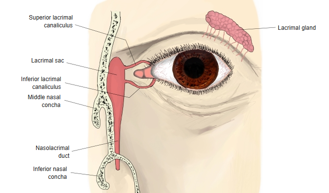
- Lacrimal apparatus
- Lacrimal gland
- Lacrimal sac
- Lens
- Macula lutea
- Nasolacrimal duct
- Optic disc
- Pupil
- Retina
- Sclera
- Vascular layer of eyeball
- Vitreous body
Parts of the visual organ
| Part | Main components | Role |
|---|---|---|
| Eyeball | Wall: fibrous, vascular and inner layers. Contents: aqueous humor, lens, vitreous body | Focuses light on the retina, where it is turned into nerve signals |
| Optic nerve | Axons of the retinal ganglion cells | Carries the visual signals from the eyeball to the brain |
| Accessory structures | Eyelids, conjunctiva, lacrimal apparatus, extraocular muscles, eyebrows | Protect, moisten and move the eyeball |
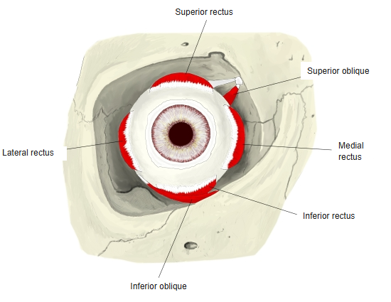
- Extraocular muscles: superior, inferior, medial and lateral rectus; superior and inferior oblique – described in the muscles section.
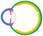
Three layers of the wall (diagram) 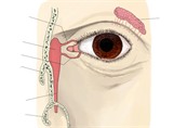
Lacrimal apparatus 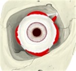
Right extraocular muscles, anterior view
References and sources
- Further reading: Visual organ, Wikipedia.