Fasciae of urogenital diaphragm (Latin: fasciae diaphragmatis urogenitalis)
urogenital
diaphragm
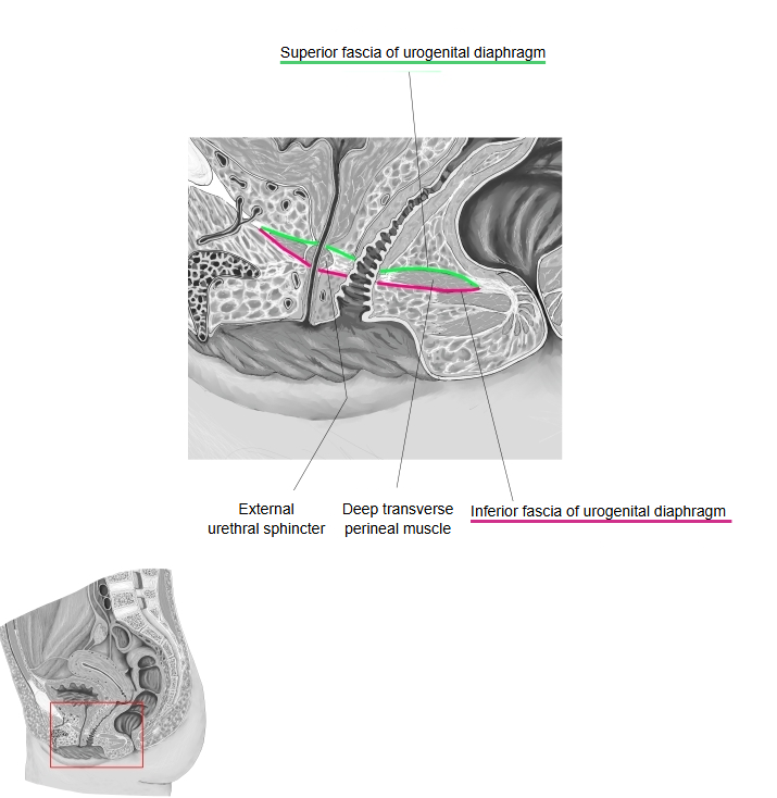
- Superior fascia of urogenital diaphragm : often not clearly defined
- Inferior fascia of urogenital diaphragm : fairly thick and strong compared with the superior fascia; also called the perineal membrane
- Note : some sources call them "internal" and "external" instead of "superior" and "inferior".
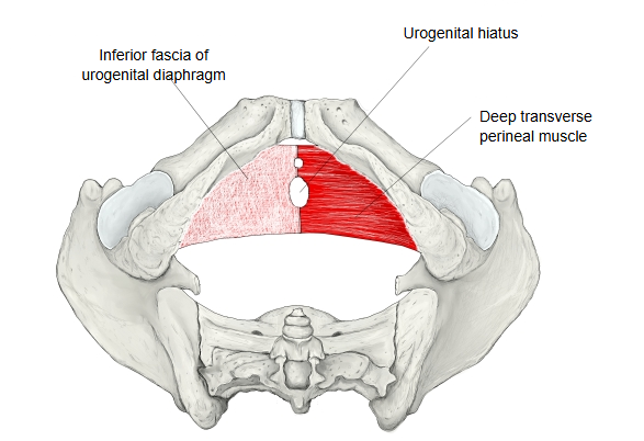
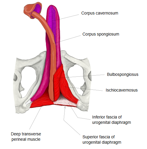
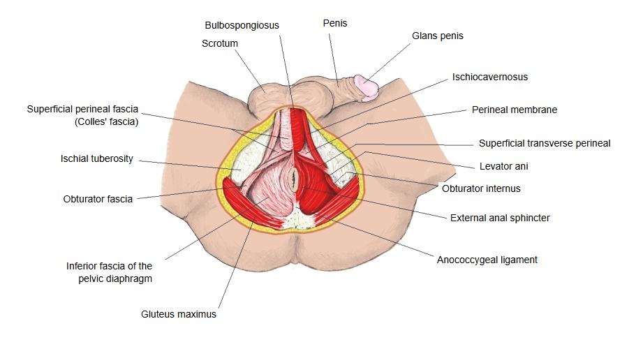
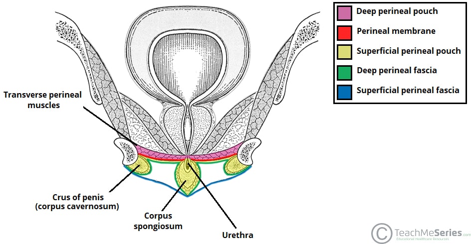
In its classical description the urogenital diaphragm is a muscular sheet stretched across the front part of the pelvic outlet, between the two ischiopubic rami (the pubic arch); its muscles lie between the two fasciae. The superior fascia is the thin upper layer facing the pelvic cavity and the levator ani of the pelvic diaphragm. The inferior fascia is attached to the ischiopubic rami; the roots of the external genitalia and the bulbospongiosus and ischiocavernosus lie below it, in the perineum, and at its back edge it joins Colles' fascia and the perineal body. Behind the muscles the two layers join each other; in front they fuse to form the transverse perineal ligament.
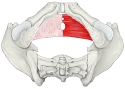 Urogenital diaphragm (inferior)
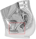 Sagittal section of pelvis
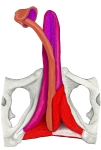 Male perineal muscles (anterior)
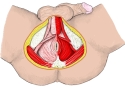 Male perineum (inferior)
Reference websites
[Wikipedia]
· Superior fascia of the urogenital diaphragm, Wikipedia.
· Urogenital diaphragm, Wikipedia.
· Perineal membrane, Wikipedia.
[Websites with illustrations]
· Fasciae of the pelvis and perineum (Gray's Anatomy, fig. 407) (Wikimedia Commons)

· Layers of the urogenital triangle in the perineum (TeachMeAnatomy)
· Urogenital diaphragm (illustration) (Pinterest)

· Perineum and urogenital diaphragm (illustration) (Pinterest)

Structure
Followed medially from obturator internus, the obturator fascia is attached by its deeper fibres to the inner margin of the pubic arch, while its superficial fibres pass over this attachment and continue as the superior fascia of the urogenital diaphragm. Behind, the superior fascia is continuous with the inferior fascia and with the superficial perineal fascia (Colles' fascia); in front, it is continuous with the fascial sheath of the prostate and fuses with the inferior fascia to form the transverse perineal ligament (transverse ligament of the pelvis).
Controversy
Some sources doubt that a separate superior fascia, or a urogenital diaphragm as a closed muscular sheet, exists at all, and describe only the perineal membrane with the deep perineal pouch above it. Even so, the layer is still used in many anatomy resources to describe how the contents of the perineum are divided.
Related terms
- Urogenital diaphragm
- Perineal membrane
- Perineal body
- Obturator fascia
- Pelvic diaphragm
- Perineal region
- Pubic arch
- Deep transverse perineal
References and sources
- Further reading: Urogenital diaphragm, Wikipedia.
- Further reading: Perineal membrane, Wikipedia.
- Federative International Programme for Anatomical Terminology (FIPAT). Terminologia Anatomica, 2nd ed. (TA2), 2019.