Perineal body (Latin: corpus perineale)
Also called the central tendon of perineum
- Overview
- Muscles that form it
- Structures attached to it
- Function
- Reference websites
- (Wikipedia, websites with illustrations)
- Related terms
Overview
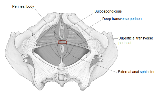
It is a thick, firm mass of fibromuscular tissue at the centre of the perineum, at the back edge of the perineal membrane, between the urogenital triangle in front and the anal triangle behind. It is made of dense connective tissue mixed with smooth and skeletal muscle fibres and acts as an anchor point for the muscles of the pelvic floor. Its position is marked on the skin by the middle of the perineal raphe. It can be stretched or torn during childbirth. Sources differ on which muscles are counted as part of it rather than as attached to it. Behind the anus, the corresponding midline tissue is the anococcygeal body.
Size
Sources differ on its exact size.
Average size: ultrasound measurements in healthy adult women who have not given birth give a height of roughly 2 cm and a front-to-back length of about 1.5–2 cm.
Muscles that form it
The perineal body is formed mainly by the following muscles.
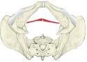
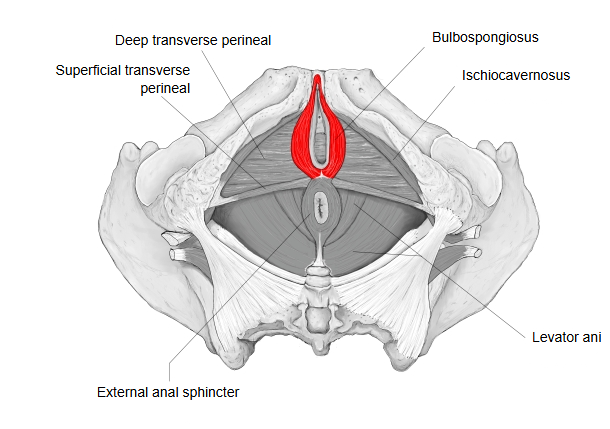
· Superficial transverse perineal: a thin muscle that runs across the pelvic floor from the ischial tuberosities.
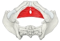
· Deep transverse perineal: a broader sheet that runs across the front half of the pelvic floor between the ischiopubic rami, below the urogenital hiatus.
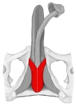
· Bulbospongiosus: a muscle in the middle of the perineum that supports urination, ejaculation and erection; it differs between the sexes. Its fibres arise from the perineal body and run forward around the bulb of the penis or the vaginal opening.
· Levator ani: of its parts, the puborectalis and the pubococcygeus send fibres into it.
 Superficial transverse perineal
 Deep transverse perineal
 Bulbospongiosus (male)
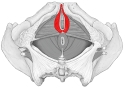 Bulbospongiosus (female)
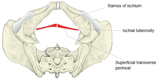
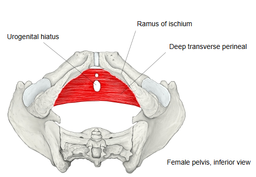
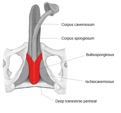
Structures attached to it
The following structures are attached to the perineal body.
1. 1. External anal sphincter: its front fibres attach firmly to the back of the perineal body, pull the anal canal forward and steady the perineal region.
2. 2. Longitudinal muscle of the rectum: some fibres of the outer, longitudinal layer of the muscle coat of the rectum reach the back of the perineal body.
3. 3. Rectovaginal septum / rectoprostatic fascia (Denonvilliers' fascia):
Female: the rectovaginal septum between the rectum and the vagina joins the top of the perineal body below.
Male: the rectoprostatic fascia joins the perineal body and separates the rectum from the prostate.
Function
1. 1. Reinforcing the pelvic floor: it helps to close the wide gap of the pelvic outlet and to hold the bladder, uterus and rectum in place.
2. 2. Control of excretion and reproduction: the muscles attached to it act from it as a fixed point, so it is a key point for holding back and for passing urine and faeces.
Reference websites
[Wikipedia]
· Perineal body, Wikipedia.
[Websites with illustrations]
· Muscles attaching to the perineal body (TeachMeAnatomy)
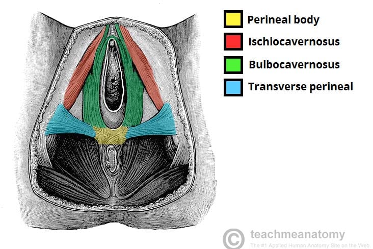
· Perineal muscles and the perineal body (inferior view) (Anatomy.app)

Related terms
- Perineal membrane
- Perineum
- Perineal region
- Anal triangle
- Pelvic diaphragm
- Anococcygeal body
- External anal sphincter
- Bulbospongiosus
- Superficial transverse perineal
References and sources
- Further reading: Perineal body, Wikipedia.
- Federative International Programme for Anatomical Terminology (FIPAT). Terminologia Anatomica, 2nd ed. (TA2), 2019.