Alveolar canals (Latin: canales alveolares)
The alveolar canals are the thin bony canals in the outer walls of the body of the maxilla that carry nerves and blood vessels; this is the collective name.
Body of the maxilla: the main part of the maxilla, apart from its four processesMainly for the upper teeth, the gums and the tooth sockets
· The outer wall of the body of the maxilla is in practice the side wall of the maxillary sinus.
There are 3 kinds of alveolar canals: the anterior and middle superior alveolar canals, which branch from the infraorbital canal, and the posterior superior alveolar canals, whose entrances open on the maxillary tuberosity.
Infraorbital canal: the continuation of the infraorbital groove in the floor of the orbitEntrances: the alveolar foramina
Alveolar
canals
canals
- Anterior superior alveolar canal : branches from the infraorbital canal (the continuation of the infraorbital groove) and runs in the front wall of the body of the maxilla.
Contents: anterior superior alveolar nerve, artery and vein, to the incisors and canine. - Middle superior alveolar canal : (not always present) branches from the infraorbital canal (some sources: from the infraorbital groove) and runs down near the junction of the front and back walls.
Contents: middle superior alveolar nerve, artery and vein, to the premolars (the course of the vessels is less clearly described). - Posterior superior alveolar canals : begin at the alveolar foramina (2–3) on the maxillary tuberosity and run forwards in the back wall.
Contents: posterior superior alveolar branches of the maxillary nerve, posterior superior alveolar artery and veins, to the molars.
Note: sources differ. Some, including Wikipedia, use "alveolar canals" only for the posterior superior alveolar canals that open on the maxillary tuberosity; others use it for all three groups.
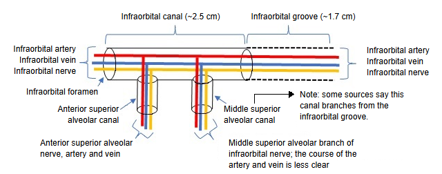
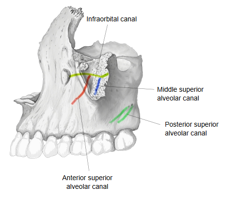
In more detail:
The alveolar canals run in the thin walls of the maxillary sinus. The anterior and middle canals branch from the infraorbital canal; the posterior canals begin at the alveolar foramina on the maxillary tuberosity. The nerves inside them, the anterior, middle and posterior superior alveolar branches of the maxillary nerve, join to form the superior dental plexus above the roots of the upper teeth.
Related terms
- Dental alveoli
- Alveolar border
- Alveolar arch
- Alveolar foramina
- Alveolar bone
- Alveolar yokes
- Alveolar crest
- Gingival sulcus
- Apical foramen
- Anterior superior alveolar canal
- Posterior superior alveolar canal
- Infraorbital canal
- Maxillary tuberosity
- Maxillary sinus
- Body of maxilla
- Maxillary nerve
References and sources
- Further reading: Maxilla, Wikipedia.
- Further reading: Superior alveolar nerves, Wikipedia.
- Federative International Programme for Anatomical Terminology (FIPAT). Terminologia Anatomica, 2nd ed. (TA2), 2019.