Infraorbital canal (Latin: canalis infraorbitalis)
· Its length varies from person to person; a figure of roughly 2.5 cm is sometimes given.
Not every description of the canal names the vein, but the infraorbital vein runs with the infraorbital artery and nerve through the infraorbital foramen, the canal and the groove, back towards the inferior orbital fissure.
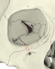 Infraorbital groove, canal and foramen
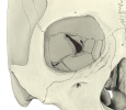 Orbit (anterior view)
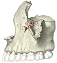 Infraorbital canal in the maxilla
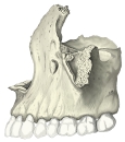 Maxilla (lateral view)
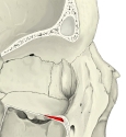 Infraorbital canal (section)
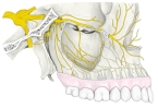 Maxillary nerve and its branches
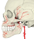 Maxillary artery and its branches
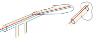 Infraorbital and alveolar canals (diagram)
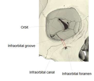
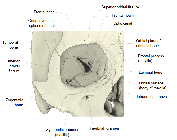
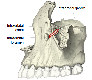
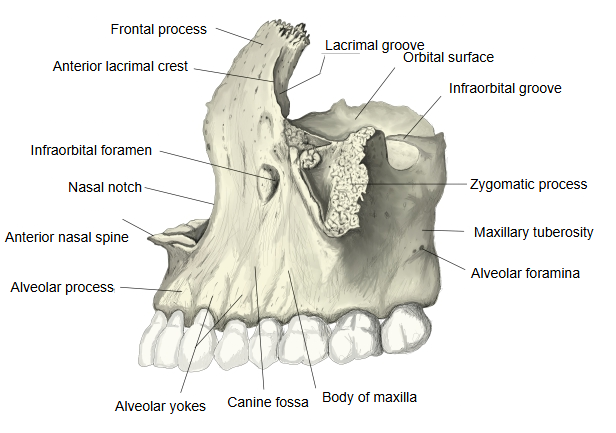
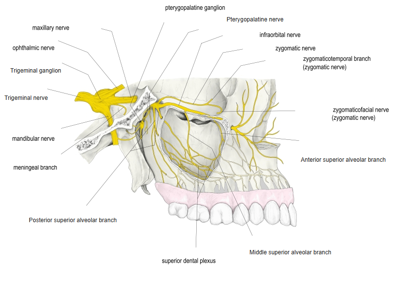
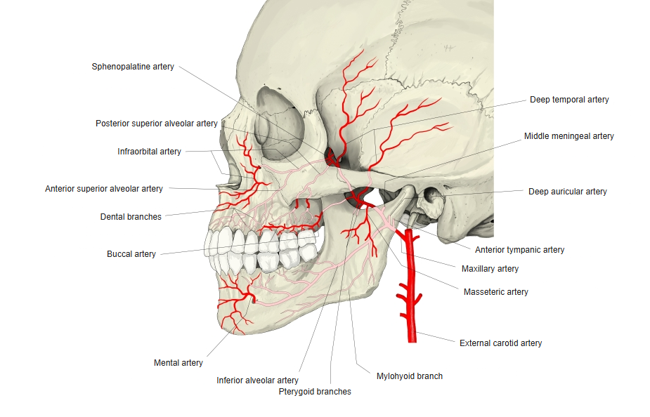
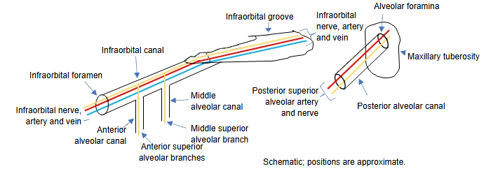
The front end of the orbital surface sinks into the body of the maxilla to become the infraorbital canal, which lies above the maxillary sinus and opens at the infraorbital foramen. The infraorbital groove and canal are the path of the infraorbital nerve, the end of the maxillary nerve, and of the infraorbital artery, a terminal branch of the maxillary artery. The infraorbital suture often runs from the start of the canal to the foramen.
Reference websites
[Wikipedia]
· Infraorbital canal, Wikipedia.
· Infraorbital nerve, Wikipedia.
[Websites with illustrations]
· Infraorbital artery (Earth's Lab)

· Infraorbital canal and nerve (figure) (Springer Nature)

· Maxilla and infraorbital canal (figure) (Elsevier)

Related terms
- Infraorbital foramen
- Body of maxilla
- Maxillary sinus
- Orbit
- Inferior orbital fissure
- Infraorbital nerve
- Infraorbital artery
References and sources
- Further reading: Infraorbital canal, Wikipedia.
- Further reading: Infraorbital nerve, Wikipedia.
- Federative International Programme for Anatomical Terminology (FIPAT). Terminologia Anatomica, 2nd ed. (TA2), 2019.