Maxillary sinus (Latin: sinus maxillaris)
Also called the antrum of Highmore
sinuses
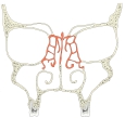
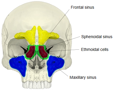
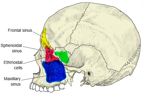
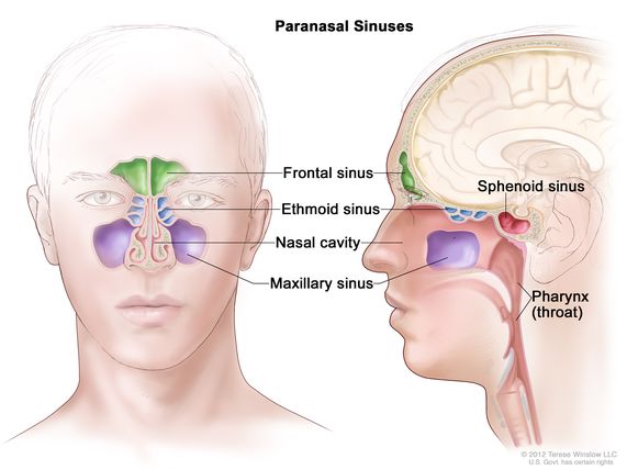
- Maxillary sinus : in the maxilla; the largest paranasal sinus.
- Frontal sinus : in the frontal bone.
- Ethmoidal cells : in the ethmoid bone; divided into many small spaces.
- Sphenoidal sinus : in the sphenoid bone; it lies behind the ethmoidal cells.
Features of each wall
| Wall | Features |
|---|---|
| Anterior wall | Thin, especially in the canine fossa; the anterior (facial) surface of the maxilla, behind the cheek. |
| Posterior wall | Thicker than the anterior wall; the infratemporal surface. |
| Roof | Thin: it is the orbital surface of the maxilla, the floor of the orbit. The infraorbital canal often raises a ridge on it. |
| Medial wall | The thinnest wall; the nasal surface, with the opening of the sinus near its top. |
| Floor | Formed by the alveolar process; its thickness varies from place to place: · over the first molar and second premolar: the thinnest; · over the second molar: next thinnest (sources differ on which tooth lies closest). The roots of these teeth often reach the sinus. The floor shows many ridges and hollows because of the septa between the tooth sockets. |
| The anterior and posterior walls show many fine alveolar grooves, alveolar canals and alveolar foramina for the superior alveolar nerves and vessels. | |
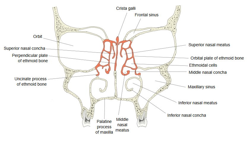
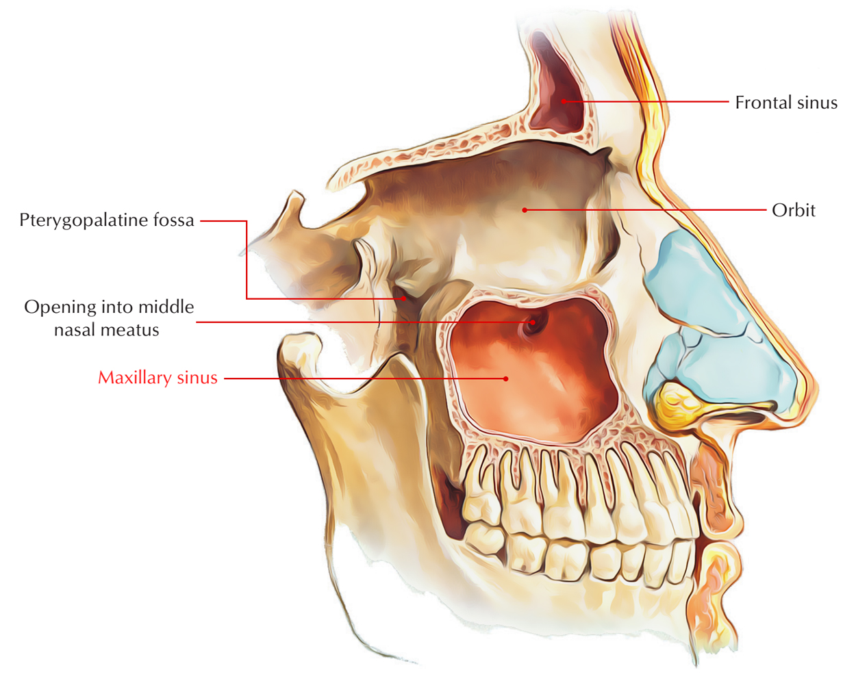
The sinus opens into the middle nasal meatus through the maxillary hiatus, which in the complete skull is narrowed by the ethmoid, the palatine bone and the inferior nasal concha to a small opening (about 2-3 mm) high on the medial wall. In adults it holds roughly 10-15 ml; its size varies a lot between people and between the two sides. It extends into small recesses: alveolar (down toward the tooth roots), zygomatic (outwards) and infraorbital (upwards); some sources also describe a palatine recess. It is lined by a thin mucous membrane with cilia that sweep mucus toward the opening.
Because the opening lies high on the medial wall, the sinus does not drain by gravity when the head is upright, and infection (maxillary sinusitis) is common. Infection of an upper molar or premolar root can spread into the sinus, and pain from the sinus can be felt in the upper teeth. Sensation comes from the superior alveolar nerves and the infraorbital nerve, branches of the maxillary nerve (V2).
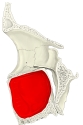 Maxilla (sagittal section)
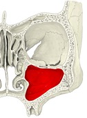 Coronal section
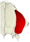 Horizontal section (from above)
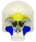 Paranasal sinuses (anterior)
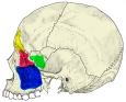 Paranasal sinuses (lateral)
 Nasal cavity (coronal section)
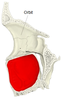
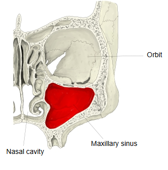
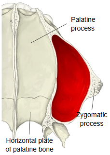
The maxillary sinus is a large cavity in the body of the maxilla and one of the paranasal sinuses. Its shape follows the body of the maxilla: roughly a pyramid with its apex pointing upwards and laterally into the zygomatic process and its base on the nasal surface, where the large, irregular maxillary hiatus lies.
Reference websites
[Wikipedia]
· Maxillary sinus, Wikipedia.
· Paranasal sinuses, Wikipedia.
[Websites with illustrations]
· Maxillary sinus (Earth's Lab)
· Paranasal sinuses (National Cancer Institute)
· Maxillary sinus (anatomy.net)

Where it is found
Related terms
- Body of maxilla
- Maxillary hiatus
- Middle nasal meatus
- Frontal sinus
- Sphenoidal sinus
- Ethmoidal air cells
- Alveolar process
References and sources
- Further reading: Maxillary sinus, Wikipedia.
- Further reading: Paranasal sinuses, Wikipedia.
- Federative International Programme for Anatomical Terminology (FIPAT). Terminologia Anatomica, 2nd ed. (TA2), 2019.