Zygomatic process (Latin: processus zygomaticus)
A process that reaches out to meet the zygomatic bone (cheekbone); the frontal bone, the temporal bone and the maxilla each have one, and each joins the zygomatic bone at a suture.
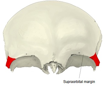
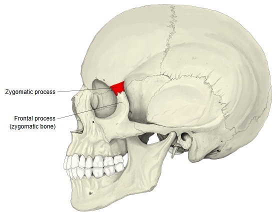
Zygomatic process of the frontal bone
The part at the lateral end of the supraorbital margin of the frontal bone that projects downwards and laterally. It joins the frontal process of the zygomatic bone at the frontozygomatic suture.
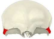 Frontal bone (anterior view)
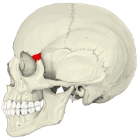 Skull (left lateral view)
Zygomatic process of the temporal bone
Zygomatic bone (temporal process) + temporal bone (zygomatic process) ⇒ zygomatic arch
· The frontal bone and the maxilla also have a process of the same name (see the other sections of this page).
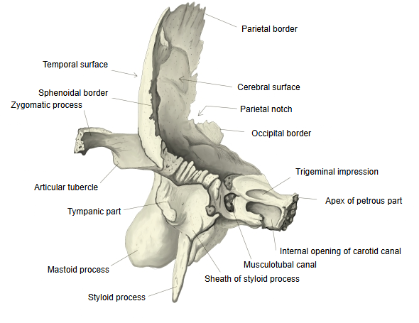
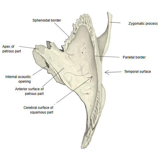
- Squamous part : occupies the middle and the whole upper part; it has two surfaces, temporal and cerebral. Related terms: groove for the middle temporal artery, zygomatic process (articular tubercle), suprameatal spine.
- Petrous part : the lower part, made up of two parts (the mastoid part, with the mastoid process, and the petrous part proper); it forms part of the cranial base.
- Tympanic part : the smallest part: a curved, half-tube-shaped thin plate that forms the front and lower walls of the external acoustic meatus.
Course
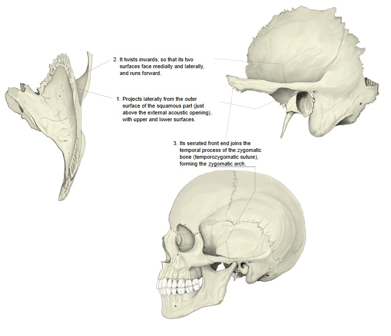
1. It projects laterally from the outer surface of the squamous part (just above the external acoustic opening), with an upper and a lower surface.
2. It then twists inwards, so that its two surfaces face medially and laterally, and runs forward.
3. Its serrated front end joins the temporal process of the zygomatic bone (the temporozygomatic suture), forming the zygomatic arch.
⇒ Illustrated explanation
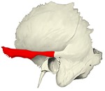 Temporal bone: lateral view
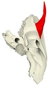 Temporal bone: medial view
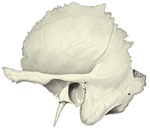 Temporal bone: lateral view, main features
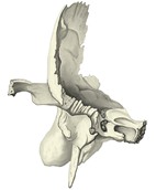 Temporal bone: anterior view
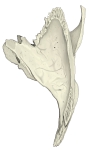 Temporal bone: superior view
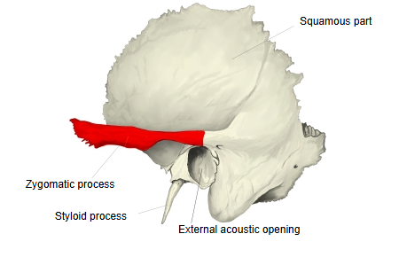
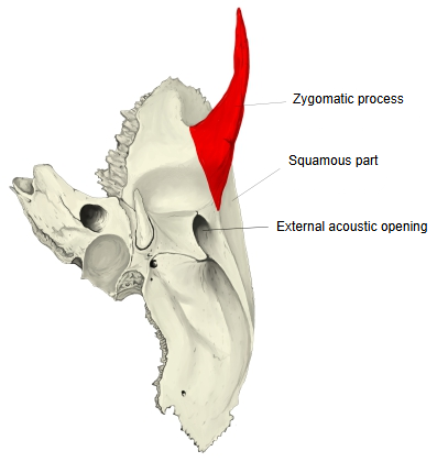
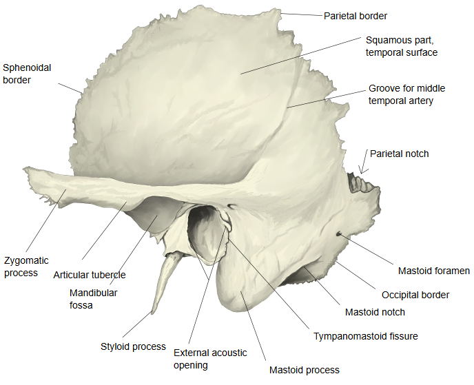
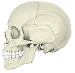 Skull (left lateral view)
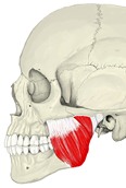 Masseter (a muscle of mastication)
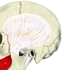 Temporal fascia
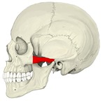 Zygomatic arch
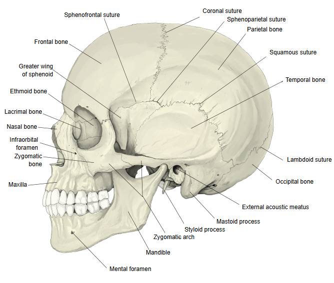
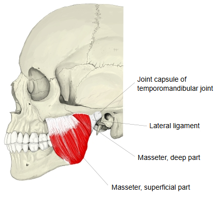
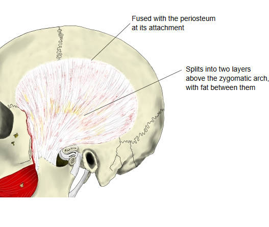
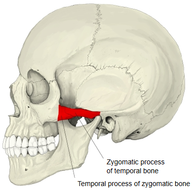
The zygomatic process of the temporal bone is a long, arched bar from the lower part of the squamous part. Its upper border is long, thin and sharp and gives attachment to the temporal fascia; its lower border is short, thick and arched and gives attachment to some fibres of masseter. Its lateral surface is convex and lies just under the skin; its medial surface is concave and also gives attachment to masseter.
Its serrated front end joins the zygomatic bone. Its back end is fixed to the squama by two roots: the posterior root, continuing the upper border, runs back above the external acoustic meatus; the anterior root, continuing the lower border, is short, broad and strong, turns medially and ends as the articular tubercle in front of the mandibular fossa, part of the temporomandibular joint.
Zygomatic process of the maxilla
- Body of maxilla : the main, roughly pyramid-shaped part of the maxilla; it contains the maxillary sinus and has four surfaces.
- Processes
- Frontal process : a long, flat process reaching upwards, between the nasal bone and the lacrimal bone.
- Zygomatic process : a thick, short process from the lateral side of the body that joins the zygomatic bone.
- Palatine process : projects almost horizontally inwards from the lower part of the body.
- Alveolar process : the downward continuation of the body, holding the sockets for the roots of the teeth.
· It joins the zygomatic bone by a rough, triangular surface (the zygomaticomaxillary suture).
· The frontal bone and the temporal bone also have a process of the same name.
Related terms
Zygomaticoalveolar crest: a strong bony ridge that runs from the lower border of the zygomatic process towards the first molar.
Infrazygomatic crest: an older name for this lower border, which runs downwards as the boundary between the anterior and infratemporal surfaces of the body of maxilla and reaches the alveolar part over the first or second molar.
The zygomatic process is a three-sided, prism-like process that runs forwards and laterally from the upper lateral corner of the body of maxilla; the rough surface at its end joins the zygomatic bone.
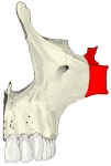 Maxilla: anterior view
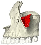 Maxilla: lateral view
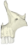 Maxilla: anterior view, main features
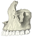 Maxilla: lateral view, main features
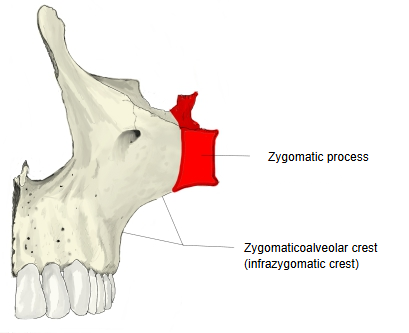
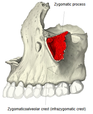
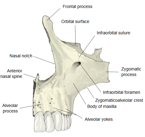
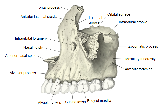
The zygomatic process of the maxilla (also called the malar process) is a rough, triangular eminence where the anterior, infratemporal and orbital surfaces of the body meet. In front it forms part of the anterior surface; behind, it is concave and forms part of the infratemporal fossa; above, it is rough and serrated for the joint with the zygomatic bone; below, it forms the prominent arched border between the anterior and infratemporal surfaces.
⇒ Back to the frontal bone page
Where it is found
Related terms
- Zygomatic arch
- Temporal process
- Squamous part
- Temporomandibular joint
- Body of maxilla
- Zygomaticomaxillary suture
- Zygomatic bone
References and sources
- Further reading: Zygomatic process of temporal bone, Wikipedia.
- Further reading: Zygomatic process of maxilla, Wikipedia.
- Federative International Programme for Anatomical Terminology (FIPAT). Terminologia Anatomica, 2nd ed. (TA2), 2019.