Inner phalangeal cell
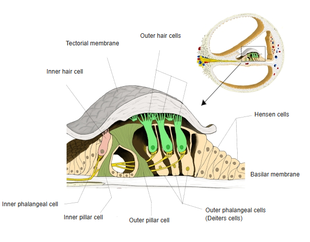
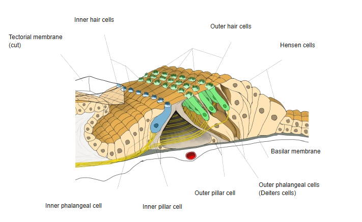
· They lie on the inner side of the inner pillar cells, in a single row along the whole length of the cochlear duct, and are the supporting cells of the inner hair cells. Each cell surrounds the base and sides of an inner hair cell, so the hair cell is held above the basilar membrane and does not touch it; its slender upper end reaches the surface and joins the heads of the pillar cells and the tops of the hair cells in the reticular membrane. They are the inner counterpart of the outer phalangeal cells (Deiters' cells); further inwards they are followed by the border cells and the cells lining the internal spiral sulcus.
of
Corti
- Hair cells
- Inner hair cells : in a single row on the inner side of the inner pillar cells, with stereocilia on their free surface; about 3,500.
- Outer hair cells : in several rows on the outer side of the outer pillar cells; about three to four times as many as the inner hair cells.
- Supporting cells
- Inner pillar cells : outside the inner hair cells; with the outer pillar cells they form the tunnel of Corti.
- Outer pillar cells : inside the outer hair cells; with the inner pillar cells they form the tunnel of Corti.
- Inner phalangeal cells : on the inner side of the inner pillar cells; they support the inner hair cells.
- Outer phalangeal cells : = cells of Deiters. Outside the outer pillar cells; they support the outer hair cells, in rows matching them. Related terms: space of Nuel, phalangeal process, reticular membrane, Retzius fibre.
- Cells of Hensen : several rows of cells outside the outer phalangeal cells, forming the outer slope of the organ of Corti.
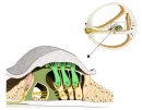 Spiral organ (section)
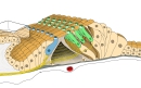 Spiral organ (3D view)
Reference websites
[Wikipedia]
· Organ of Corti, Wikipedia.
[Websites with illustrations]
· Organ of Corti (diagram) (Wikimedia Commons)

Related terms
- Inner hair cell
- Organ of Corti
- Outer phalangeal cell
- Outer pillar cell
- Outer hair cell
- Reticular membrane
- Phalangeal process
- Basilar membrane
References and sources
- Further reading: Organ of Corti, Wikipedia.
- Federative International Programme for Anatomical Terminology (FIPAT). Terminologia Anatomica, 2nd ed. (TA2), 2019.