Space of Nuel (Latin: cuniculus medius)
Also called Nuel's space or the middle tunnel
· It lies next to (just outside, lateral to) the tunnel of Corti.
· It is filled with fluid. As it lies inside the cochlear duct, one might expect this fluid to be endolymph, like that of the cochlear duct, but it is not:
the reticular membrane at the top of the organ of Corti separates the two fluids.
The sensory hair cells have only their apical surface, which carries the stereocilia, in the endolymph; their cell bodies are bathed in a fluid like perilymph (cortilymph), which also fills the space of Nuel and the tunnel of Corti.
of
Corti
- Hair cells
- Inner hair cells : in a single row on the inner side of the inner pillar cells, with stereocilia on their free surface.
- Outer hair cells : in several rows on the outer side of the outer pillar cells; several times as many as the inner hair cells.
- Supporting cells
- Inner pillar cells : outside the inner hair cells; with the outer pillar cells they form the tunnel of Corti.
- Outer pillar cells : inside the outer hair cells; with the inner pillar cells they form the tunnel of Corti.
- Inner phalangeal cells : on the inner side of the inner pillar cells; they support the inner hair cells.
- Outer phalangeal cells : = cells of Deiters. Outside the outer pillar cells; they support the outer hair cells in several rows. Related terms: space of Nuel, phalangeal process, reticular membrane, Retzius fibre.
- Cells of Hensen : several rows of cells outside the outer phalangeal cells, forming the outer slope of the organ of Corti.
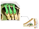 Around the outer hair cells
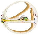 Spiral canal of the cochlea (section)
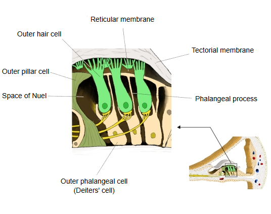
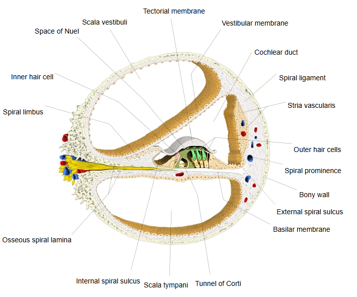
The space of Nuel is a fluid-filled space between the outer pillar cells and the innermost row of outer hair cells, which runs the whole length of the organ of Corti from the base of the cochlea to the apex. Its walls are the outer pillar cells, the outer hair cells and the outer phalangeal cells (cells of Deiters); the reticular membrane closes it from above. It opens into the tunnel of Corti through the gaps between the outer pillar cells, and nerve fibres to the outer hair cells cross it. It is named after the Belgian physician Jean-Pierre Nuel.
Reference websites
[Wikipedia]
· Organ of Corti, Wikipedia.
· Deiters cell, Wikipedia.
[Websites with illustrations]
· Organ of Corti with the tunnel of Corti and the space of Nuel (diagram) (Semantic Scholar)

· Organ of Corti, histological drawing with the spaces of Nuel (Wiley Online Library)

Related terms
- Endolymph
- Tunnel of Corti
- Organ of Corti
- Outer phalangeal cell
- Reticular membrane
- Basilar membrane
- Scala media
- Perilymph
References and sources
- Further reading: Organ of Corti, Wikipedia.
- Further reading: Deiters cell, Wikipedia.
- Federative International Programme for Anatomical Terminology (FIPAT). Terminologia Anatomica, 2nd ed. (TA2), 2019.