Intestinal epithelial cells
Also called Intestinal epithelium
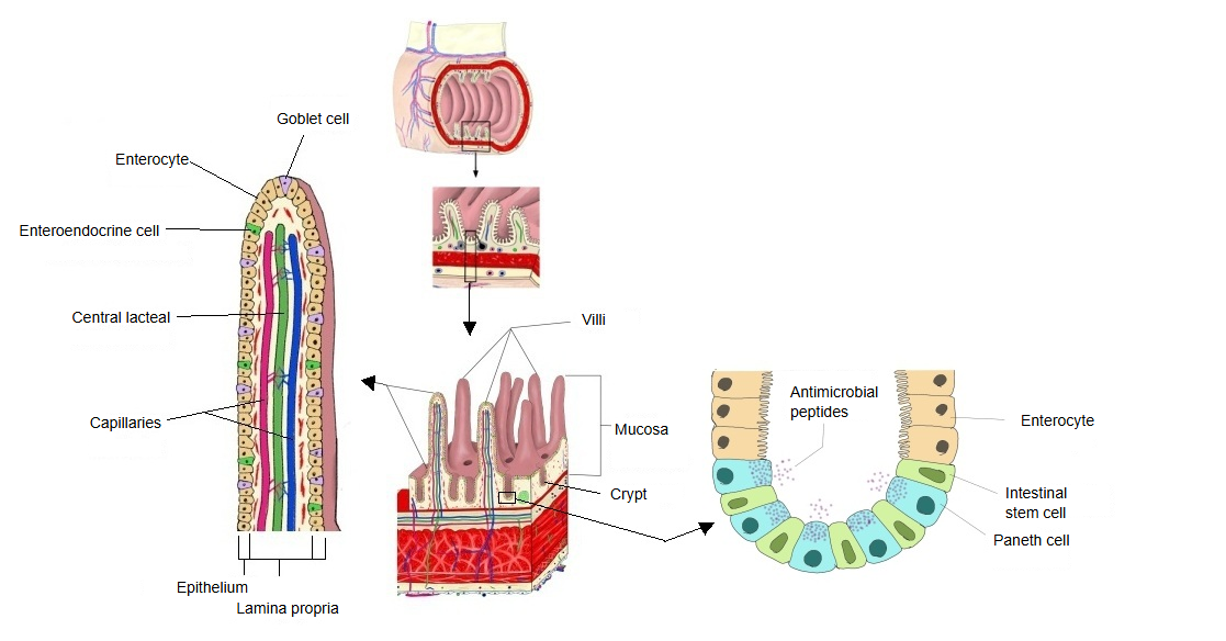
Intestinal epithelial cells is the collective name for the following five kinds of cells that make up the epithelium (a single layer of columnar cells) covering the mucosa of the intestine.
The cells of the single-layered (simple columnar) epithelium of the intestineIn the small intestine the surface is almost entirely projecting villi and sunken crypts, with very little flat surface in between
| Cell type | Main function | Location | Share (approx.) | |
|---|---|---|---|---|
| 1 | Enterocytes (absorptive cells) | Absorption of nutrients through a brush border of microvilli; brush-border digestive enzymes | Villi and crypts | about 80–90% |
| 2 | Goblet cells | Secrete mucus | Villi and crypts | about 10–15% (varies by region; more towards the colon) |
| 3 | Enteroendocrine cells | Secrete hormones | Mainly the crypts | about 1–2% |
| 4 | Paneth cells | Secrete antimicrobial peptides | Base of the crypts | about 1–3% (mainly in the jejunum and ileum) |
| 5 | Stem cells | Renew the whole epithelium every few days; the new cells move up towards the villus tips and are shed | Near the base of the crypts | very few (<1%) |
The proportions in the table are approximate and vary along the gut; sources differ. Other, less common types include tuft cells and M cells (over lymphoid nodules).
Related terms
References and sources
- Further reading: Intestinal epithelium, Wikipedia.
- Federative International Programme for Anatomical Terminology (FIPAT). Terminologia Anatomica, 2nd ed. (TA2), 2019.