Maculae cribrosae (Latin: maculae cribrosae)
Also called the cribriform areas (of the vestibule)
medial wall of
the vestibule
for nerves
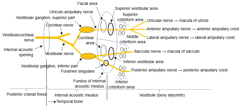
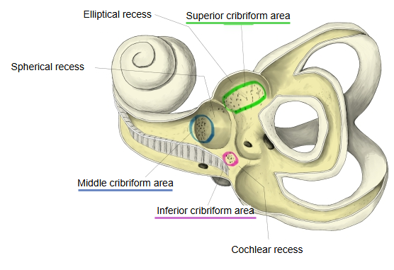
- Superior : Site: the pyramid of the vestibule and the adjacent elliptical recess.
Nerve: carries the utriculo-ampullary nerve to the superior vestibular area. - Middle : Site: the floor of the spherical recess.
Nerve: carries the saccular nerve to the inferior vestibular area. - Inferior : Site: behind the cochlear recess, at the opening of the bony ampulla of the posterior semicircular canal.
Nerve: carries the posterior ampullary nerve to the foramen singulare.
 Recesses and cribriform areas
 Left bony labyrinth, opened from the lateral side
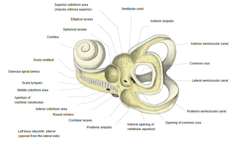
The holes pass the filaments of the vestibular nerve, the vestibular part of the vestibulocochlear nerve, between the fundus of the internal acoustic meatus and the sensory areas of the inner ear (the macula of the utricle, the macula of the saccule and the ampullary crests); the utricular nerve, for example, passes through the superior patch to the macula of the utricle. The wall they pierce, part of the bony labyrinth, separates the vestibule from the internal acoustic meatus. Sources differ on the names: the same patches are also called the superior, middle and inferior cribriform areas.
Reference websites
[Wikipedia]
· Bony labyrinth, Wikipedia.
· Vestibule of the ear, Wikipedia.
Related terms
- Vestibule
- Spherical recess
- Elliptical recess
- Cochlear recess
- Inferior cribriform area
- Fundus of internal acoustic meatus
- Vestibulocochlear nerve
References and sources
- Further reading: Bony labyrinth, Wikipedia.
- Further reading: Vestibule of the ear, Wikipedia.
- Federative International Programme for Anatomical Terminology (FIPAT). Terminologia Anatomica, 2nd ed. (TA2), 2019.