Cochlear recess (Latin: recessus cochlearis)
· It holds the lower (vestibular) end of the cochlear duct, the blind vestibular caecum.
· The vestibular crest divides below into two limbs that enclose this small depression.
of the
medial wall
of the
vestibule
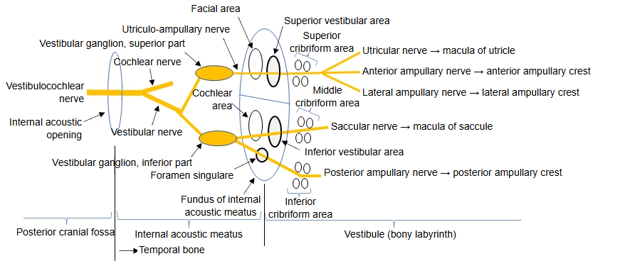
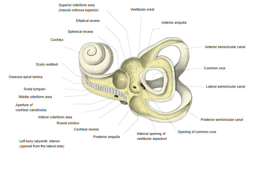
- Elliptical recess : a large, rounded recess that holds the utricle. The superior cribriform area opens here for the utriculo-ampullary nerve.
- Vestibular crest : the ridge between the elliptical and spherical recesses.
- Spherical recess : the recess that holds the saccule. The middle cribriform area opens here for the saccular nerve.
- Cochlear recess : below the spherical recess; holds the lower end of the cochlear duct. Some sources place the inferior cribriform area here, for the posterior ampullary nerve.
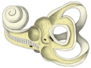 Left bony labyrinth, opened from the lateral side
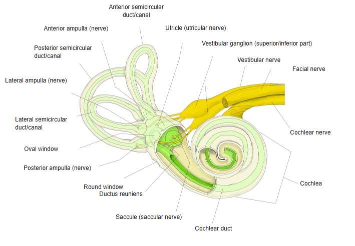
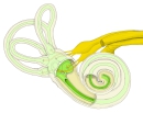 Bony and membranous labyrinth with nerves
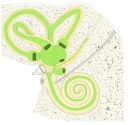 Bony and membranous labyrinth
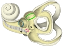 Cribriform areas and recesses
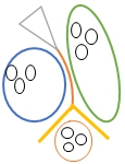 Recesses and vestibular crest (model)
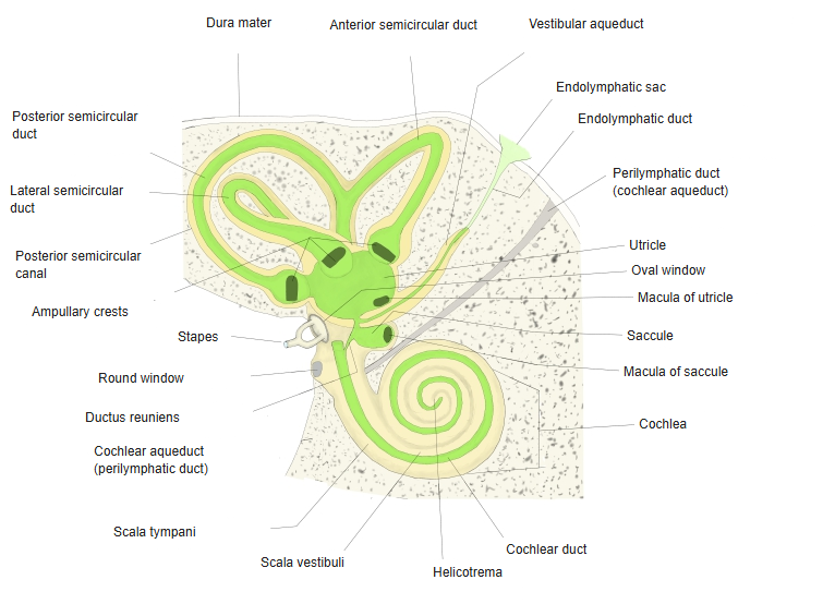
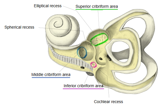
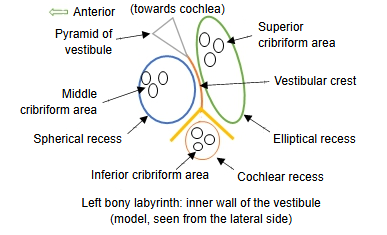
In more detail:
The vestibular crest divides below to enclose the cochlear recess, whose floor is perforated by a number of small holes for nerve fibres from the internal acoustic meatus. Sources differ on these fibres: classical texts describe fibres of the cochlear nerve to the vestibular end of the cochlear duct, while some descriptions place the inferior cribriform area here, for fibres of the posterior ampullary nerve. The medial wall of the vestibule thus has three depressions: the elliptical recess (for the utricle) above and behind, the spherical recess (for the saccule) in front, and the small cochlear recess below.
Reference websites
[Wikipedia]
· Bony labyrinth, Wikipedia.
[Websites with illustrations]
· Interior of the right osseous labyrinth (Gray's Anatomy) (Wikimedia Commons)

Related terms
References and sources
- Further reading: Bony labyrinth, Wikipedia.
- Federative International Programme for Anatomical Terminology (FIPAT). Terminologia Anatomica, 2nd ed. (TA2), 2019.