Middle talar articular surface (Latin: facies articularis talaris media calcanei)
Also called Middle facet for talus
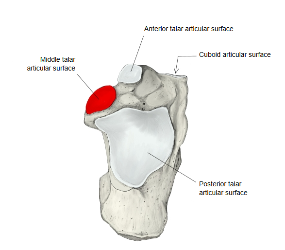
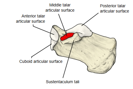
One of the articular surfaces on the upper side of the calcaneus: a small, oval surface on the upper side of the sustentaculum tali. It faces the middle calcaneal articular surface of the talus and forms part of the talocalcaneonavicular joint.
- Front surface : Cuboid articular surface
- Upper surface : Anterior talar articular surface · Middle talar articular surface · Posterior talar articular surface
Together with the anterior talar articular surface and the navicular, it makes the socket of the talocalcaneonavicular joint. It is often continuous with the anterior talar articular surface; in other feet the two are separate. The calcaneal sulcus separates it from the large posterior talar articular surface; with the sulcus tali above, this groove forms the tarsal sinus. Sources differ on whether the joint at the anterior and middle facets is counted as part of the subtalar joint or only of the talocalcaneonavicular joint.
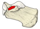 Right calcaneus (medial)
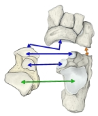 Joints of the calcaneus
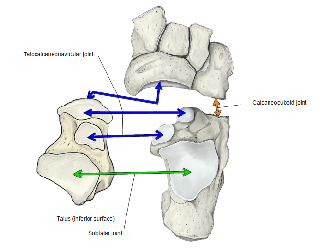
Reference websites
[Wikipedia]
· Calcaneus, Wikipedia.
· Talocalcaneonavicular joint, Wikipedia.
[Websites with illustrations]
· Bones of the right foot, dorsal surface (Gray's Anatomy, fig. 268) (Wikimedia Commons)

· Calcaneus: anatomy and pathology (Kenhub)
· Bones of the foot: tarsals, metatarsals and phalanges (TeachMeAnatomy)
Where it is found
Related terms
- Anterior talar articular surface
- Posterior talar articular surface
- Middle calcaneal articular surface
- Sustentaculum tali
- Calcaneal sulcus
- Tarsal sinus
- Talocalcaneonavicular joint
References and sources
- Further reading: Calcaneus, Wikipedia.
- Further reading: Talocalcaneonavicular joint, Wikipedia.
- Federative International Programme for Anatomical Terminology (FIPAT). Terminologia Anatomica, 2nd ed. (TA2), 2019.