Floor of mouth
Also called Floor of oral cavity
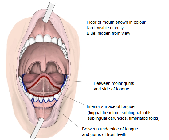
· In clinical use the floor of the mouth is the hollow, horseshoe-shaped area between the tongue and the gums of the lower teeth.
cavity
- Lips : the front wall of the mouth: the upper lip and lower lip, joined at each corner (the labial commissure).
- Oral cavity : the space inside the lips and cheeks, in two parts: the oral vestibule in front (narrow) and the oral cavity proper behind.
- Palate : the upper wall (roof) of the mouth, in two parts: the hard palate in front (the larger part) and the soft palate behind.
- Floor of mouth : the bottom of the mouth, covered by soft mucous membrane with much loose connective tissue beneath.
- Cheeks : the side walls of the mouth; they form the oral vestibule with the lips and are made of skin, muscle (buccinator) and mucous membrane.
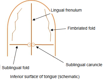
In the midline the lingual frenulum divides the floor of the mouth into right and left halves. On each side of the frenulum lie the sublingual caruncle, where the submandibular duct opens, and the sublingual fold over the sublingual gland.
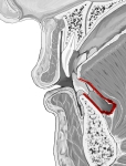 Floor of mouth (median section)
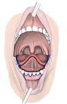 Oral cavity from the front
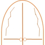 Inferior surface of tongue (schematic)
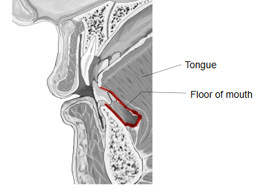
Muscles of the floor of the mouth
Some sources list the following four muscles as forming the floor of the mouth.
1. 1. Mylohyoid 2. Geniohyoid 3. Digastric 4. Stylohyoid
Other sources (for example ScienceDirect Topics) name these three:
1. 1. Mylohyoid 2. Genioglossus 3. Geniohyoid
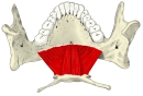 Mylohyoid and geniohyoid
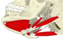 Digastric
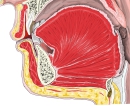 Oral cavity (median section)
Boundaries: in front and at the sides the lower gums, medially the tongue, and behind the place where the palatoglossal arch (anterior tonsillar pillar) meets the tongue. Its muscular base is formed mainly by the mylohyoid, the oral diaphragm, with the geniohyoid and genioglossus above it. The lingual frenulum divides it into right and left halves, and the ducts of the submandibular and sublingual glands open on it.
The sublingual glands lie just beneath its mucosa, raising the sublingual folds. The submandibular duct (about 5 cm long) runs forward between the sublingual gland and the genioglossus and opens at the sublingual caruncle beside the frenulum. The mucosa receives sensory fibres from the lingual nerve; the muscles are supplied by the nerve to mylohyoid (mandibular nerve), by C1 fibres running with the hypoglossal nerve (geniohyoid) and by the hypoglossal nerve (genioglossus). Lymph drains first to the submandibular and upper deep cervical (jugulodigastric) nodes, rarely to the submental nodes, and then to the lower deep cervical nodes.
Related terms
- Oral diaphragm
- Lingual frenulum
- Sublingual fold
- Sublingual caruncle
- Sublingual gland
- Mylohyoid
- Geniohyoid
References and sources
- Further reading: Floor of mouth, Wikipedia.
- Federative International Programme for Anatomical Terminology (FIPAT). Terminologia Anatomica, 2nd ed. (TA2), 2019.