Palatovaginal canal (Latin: canalis palatovaginalis)
Also called the pharyngeal canal
· A vein is usually not described in the canal. Some textbooks call the artery a branch of the sphenopalatine artery, itself the terminal branch of the maxillary artery.
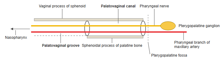
The fine groove on the under surface of the vaginal process, the palatovaginal groove, becomes deeper from back to front and is closed below by the sphenoidal process of the palatine bone to form the canal. The canal lies below and medial to the pterygoid canal; its front opening is in the lower back part of the pterygopalatine fossa, and its back opening is on the roof of the nasopharynx near the root of the pterygoid process.
 Canals under the sphenoid (front view)
 Canals under the sphenoid (from below)
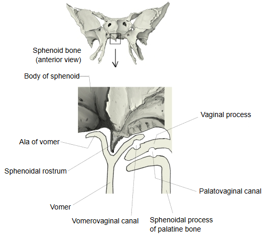
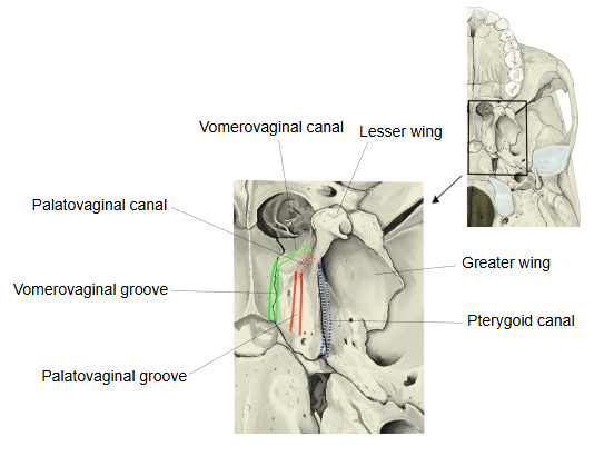
The exact positions of the palatovaginal and vomerovaginal canals in these figures are approximate.
An inconstant vomerovaginal canal may lie medial to it, between the ala of the vomer and the vaginal process, and may open into its front end. Some textbooks describe the palatovaginal canal itself as formed with the ala of the vomer and say it carries a branch of the sphenopalatine artery and branches of the pterygopalatine ganglion; sources differ.
Reference websites
[Wikipedia]
· Palatovaginal canal, Wikipedia.
· Pterygopalatine fossa, Wikipedia.
[Websites with illustrations]
· Sphenoid bone, anterior and inferior surfaces (Gray's Anatomy) (Wikimedia Commons)

· Base of the skull, outer surface (Gray's Anatomy) (Wikimedia Commons)

Related terms
- Sphenoidal process
- Ala of vomer
- Pterygopalatine fossa
- Nasopharynx
- Pharyngeal branch of maxillary artery
- Sphenoid bone
- Palatine bone
References and sources
- Further reading: Palatovaginal canal, Wikipedia.
- Further reading: Pterygopalatine fossa, Wikipedia.
- Federative International Programme for Anatomical Terminology (FIPAT). Terminologia Anatomica, 2nd ed. (TA2), 2019.