Ischial spine (Latin: spina ischiadica)
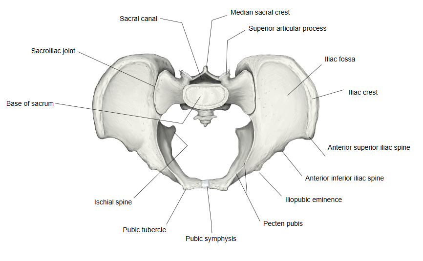
It is a pointed, triangular projection of the ischium that separates the greater sciatic notch above from the lesser sciatic notch below; its size varies between people. With the sacrotuberous ligament, the sacrospinous ligament attached to its tip turns the two notches into the greater and lesser sciatic foramina. The pudendal nerve and the internal pudendal vessels cross just behind it, on the sacrospinous ligament. The distance between the two spines is the narrowest side-to-side width of the mid-pelvis, which matters in childbirth. The spine can be felt through the vaginal wall and is used as a landmark for a pudendal nerve block and for judging how far the baby's head has descended during labour.
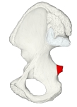 Hip bone (medial view)
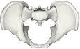 Pelvis (superior view)
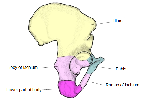
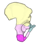 Parts of the ischium
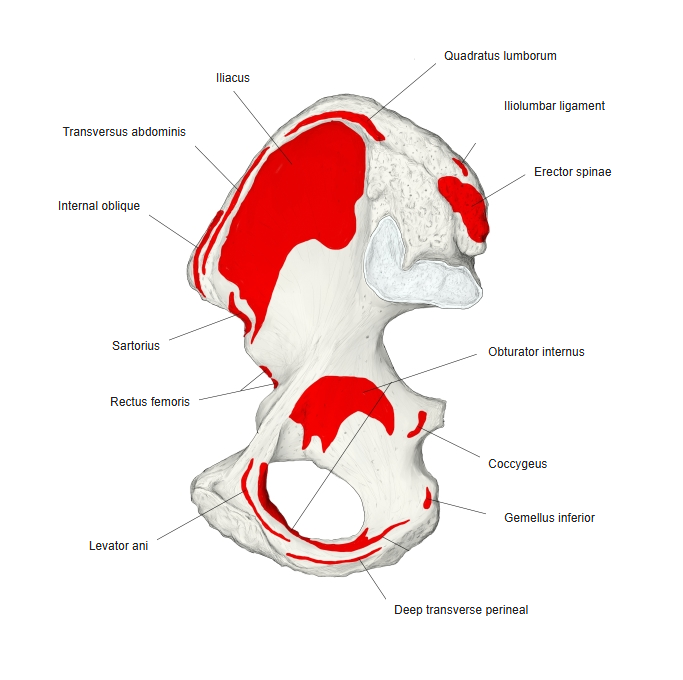
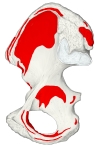 Muscle attachments (medial)
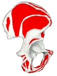 Muscle attachments (lateral)
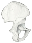 Hip bone (lateral view)
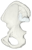 Hip bone (medial view)
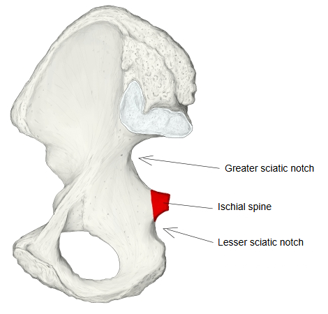
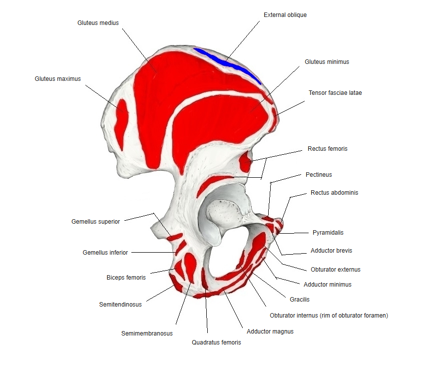
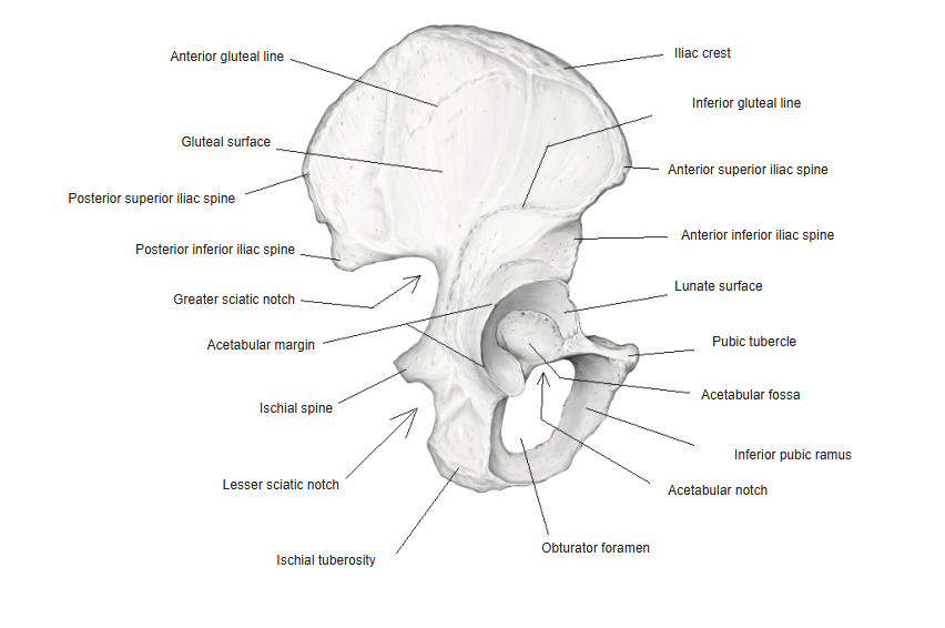
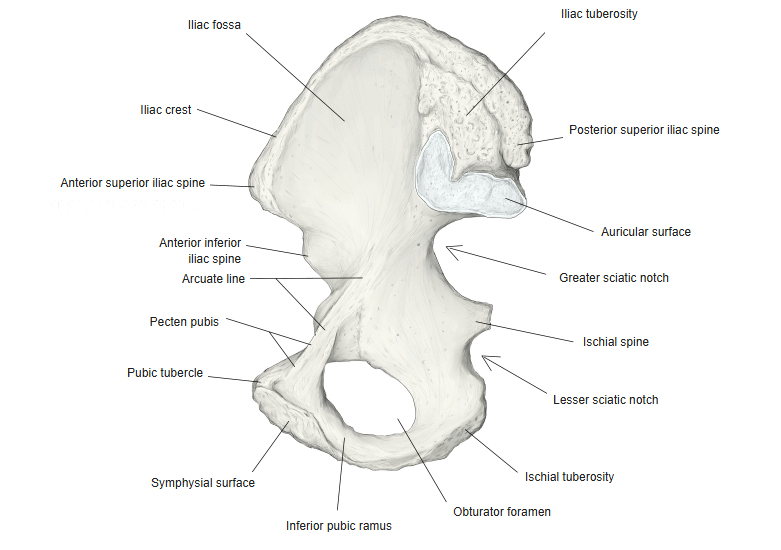
| External (gluteal) surface | gives origin to the gemellus superior |
| Internal (pelvic) surface | gives attachment to the coccygeus, the back part of the levator ani and the pelvic fascia |
| Tip | the sacrospinous ligament is attached |
Reference websites
[Wikipedia]
· Ischial spine, Wikipedia.
· Ischium, Wikipedia.
[Websites with illustrations]
· Ischial spine, medial view of the right hip bone (image) (Wikimedia Commons)

· Right hip bone, external surface (Gray's Anatomy, fig. 235) (Wikimedia Commons)

Where it is found
Related terms
References and sources
- Further reading: Ischial spine, Wikipedia.
- Further reading: Ischium, Wikipedia.
- Federative International Programme for Anatomical Terminology (FIPAT). Terminologia Anatomica, 2nd ed. (TA2), 2019.