Perirenal fat capsule (Latin: Capsula adiposa renis)
Also called the perirenal fat or adipose capsule of the kidney
- Overview
- Structures in the three coverings
- Reference websites
- (Wikipedia, websites with illustrations)
Overview
- Renal capsule : the innermost covering: a tough fibrous membrane stuck to the surface of the kidney (= fibrous capsule). At the renal hilum it continues into the renal sinus.
- Perirenal fat capsule : a layer of fat outside the renal capsule that cushions the kidney. It also surrounds the suprarenal gland, but the kidney and the suprarenal gland are wrapped separately.
- Renal fascia : a thin, strong membrane that encloses the kidney, the suprarenal gland and the perirenal fat capsule together. It has an anterior layer (= Gerota's fascia) and a posterior layer (= Zuckerkandl's fascia).
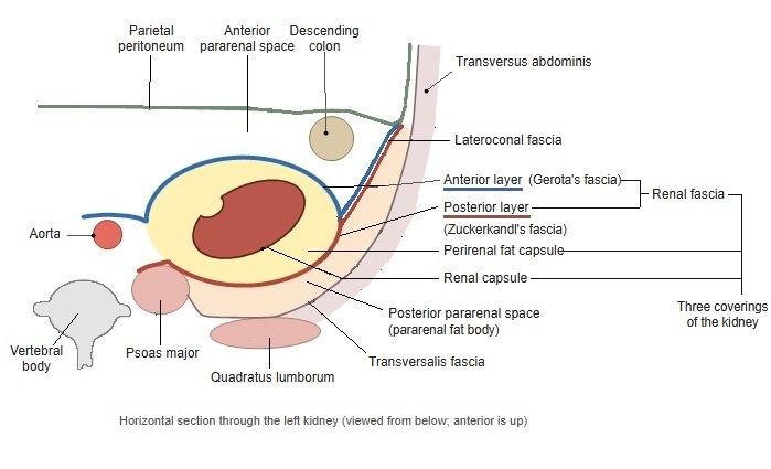
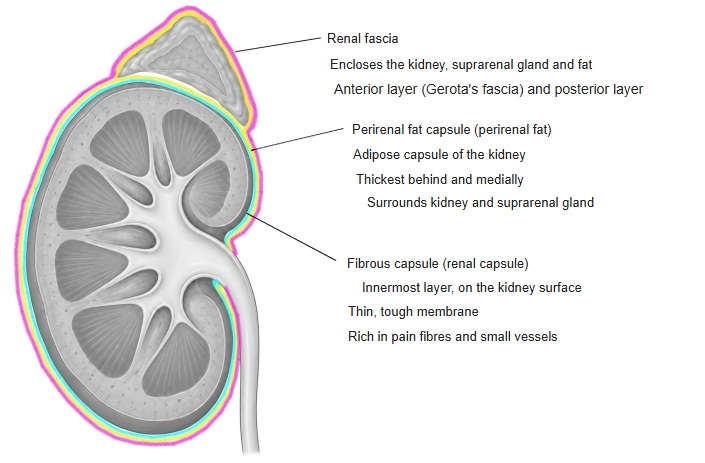
· The perirenal fat capsule is one of the coverings of the kidney: the layer of fat around the fibrous capsule on the surface of the kidney (the renal capsule). It lies inside the renal fascia. It is also called the fat capsule, the perirenal fat or the adipose capsule of the kidney.
· It is the fatty layer around the outside of the fibrous capsule, and it is best developed behind and on the medial side of the kidney.
· It encloses the suprarenal gland as well as the kidney, and its thickness changes with the state of nutrition.
· Thin bundles of connective tissue (trabeculae) cross the fat and tie the renal capsule to the renal fascia.
· The perirenal fat capsule is wrapped by the renal fascia; outside the fascia lies a separate layer of fat, the pararenal fat body. The perirenal fat cushions the kidney against knocks and helps to hold it in place.
· At the renal hilum the perirenal fat continues into the fat of the renal sinus.
Structures in the three coverings
The nerves, blood vessels, lymphatics and other structures found in the three coverings are summarised below.
| Renal capsule (fibrous capsule) | Perirenal fat capsule | Renal fascia | |
|---|---|---|---|
| Layer | Thin, strong fibrous membrane on the surface of the kidney | Fat around the kidney and the suprarenal gland | Connective-tissue membrane that encloses the perirenal fat capsule from outside (anterior and posterior layers) |
| Arteries | Fine arterial network under the capsule (from the ends of the interlobular arteries and from the capsular arteries outside the capsule) | Renal artery; suprarenal arteries (superior, middle and inferior); capsular arteries (a network in the fat) | No named arteries, veins, lymphatics or nerves (supplied by small vessels) |
| Veins | Stellate veins under the capsule (draining into the interlobular veins, also connected with the capsular veins) | Renal vein; suprarenal vein; capsular veins | |
| Lymphatics | Lymphatic network under the capsule (continuing into the lymphatics of the perirenal fat capsule) | Renal lymphatics (from the hilum along the renal vein to the lumbar lymph nodes); lymphatics from the capsule | |
| Nerves | Fine fibres from the renal plexus, many of them pain fibres (pain when the capsule is stretched) | Renal plexus (network around the renal artery); nerves to the suprarenal gland (from the greater splanchnic nerve and others, to the suprarenal medulla) | |
| Other | – | Renal pelvis and upper ureter; connective-tissue bundles (trabeculae) | Passing through the sac: renal artery and vein (medially) and the ureter (the lower end is not closed) |
Reference websites
[Wikipedia]
· Renal fascia, Wikipedia.
[Websites with illustrations]
· The kidneys (TeachMeAnatomy)
· Radiology of the perinephric space (Articl)
· Kidney layers (3D animation still) (Wikimedia Commons)

· Perirenal space on imaging (poster) (European Society of Radiology (EPOS))