Vastoadductor membrane (Latin: membrana vastoadductoria)
Also called Vastoadductor lamina or the subsartorial fascia
· It is also called the vastoadductor lamina or the subsartorial fascia.
· It forms part of the wall of the tendinous adductor canal.
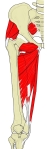 Adductor muscles and adductor hiatus
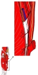 Vastoadductor membrane
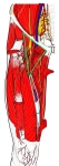 Right thigh (front)
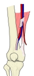 Adductor hiatus
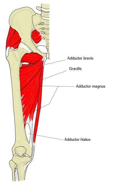
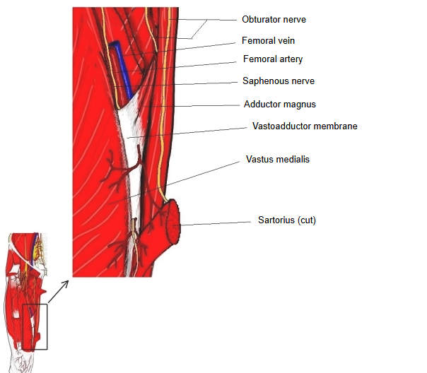
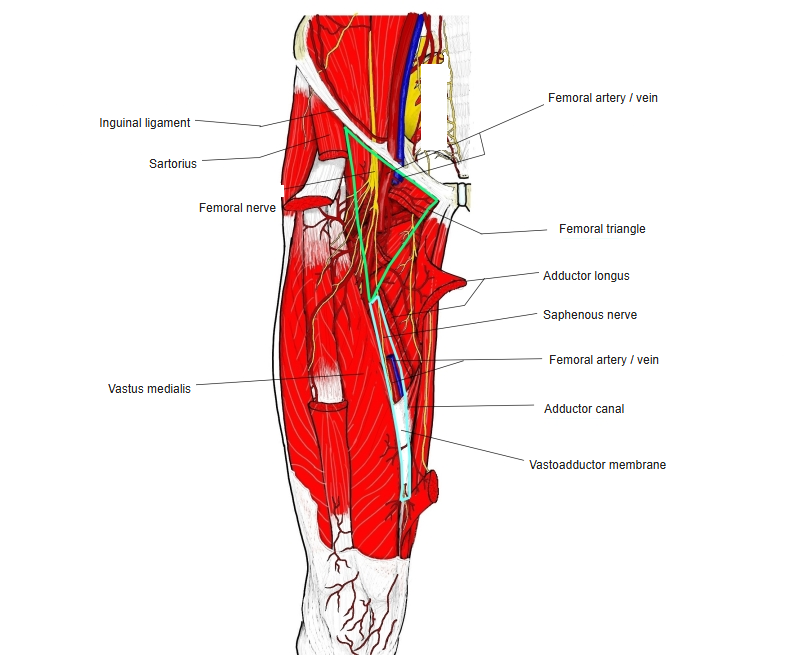
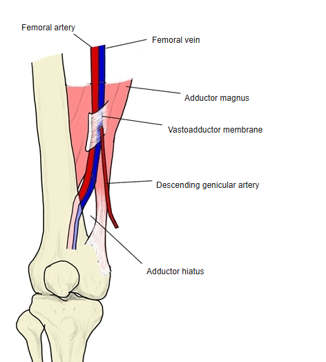
Below the iliopectineal fossa lies the muscular adductor canal, bounded by adductor longus, vastus medialis and sartorius; its lower continuation is the tendinous adductor canal. Where adductor magnus inserts on the linea aspera of the femur, a long aponeurosis runs down from its front to the medial side of vastus medialis: this is the vastoadductor membrane, and the canal between the two muscles is the tendinous adductor canal. At the lower end the insertion tendon of adductor magnus forms a tendinous arch, leaving the adductor hiatus between it and the femur. The femoral artery and vein pass from the iliopectineal fossa through the adductor canal to the popliteal fossa.
The membrane forms the roof of the adductor canal, stretching from adductor longus and adductor magnus to vastus medialis. It is strongest in the lower part of the canal, where it arises mainly from the front of the adductor magnus tendon. Sartorius lies on its surface. Its lower edge ends near the adductor hiatus, through which the femoral vessels pass into the popliteal fossa. The saphenous nerve and the descending genicular artery pierce it near its lower end.
Reference websites
[Wikipedia]
· Adductor canal, Wikipedia.
· Adductor hiatus, Wikipedia.
Related terms
- Adductor canal
- Tendinous adductor canal
- Muscular adductor canal
- Femoral triangle
- Vastus medialis
- Adductor magnus
- Saphenous nerve
References and sources
- Further reading: Adductor canal, Wikipedia.
- Further reading: Adductor hiatus, Wikipedia.
- Federative International Programme for Anatomical Terminology (FIPAT). Terminologia Anatomica, 2nd ed. (TA2), 2019.