Orbital surface of zygomatic bone (Latin: facies orbitalis ossis zygomatici)
One of the three surfaces of the body of the zygomatic bone; it forms part of the lateral wall of the orbit.
bone
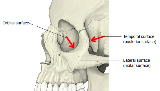
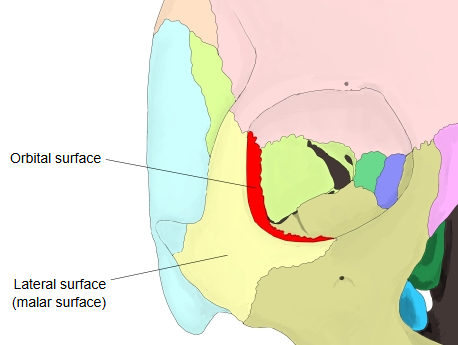
- Lateral surface : the surface that bulges outwards (= malar surface).
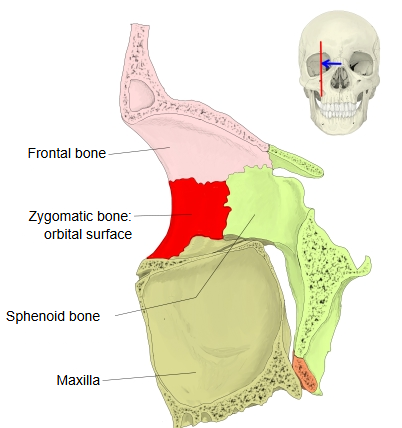
- Orbital surface : the surface that forms the lateral wall of the orbit.
- Temporal surface : the surface that forms the front wall of the temporal fossa (= posterior surface).
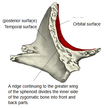
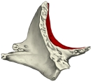 Left zygomatic bone (medial surface)
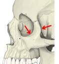 Left orbit (from the lateral side)
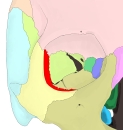 Right orbit (anterior view)
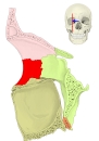 Right orbit (lateral wall)
More details
Anatomy textbooks describe it as follows.
It is a crescent-shaped surface forming part of the lateral wall of the orbit. It meets the lateral surface at a blunt, rounded margin; below, this margin continues into the orbital margin of the maxilla and above into that of the frontal bone.
In more detail:
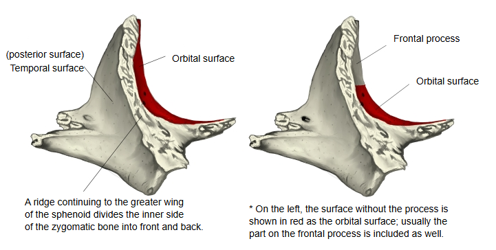
The inner side of the zygomatic bone is divided into a front and a back part by a ridge that continues to the greater wing of the sphenoid bone. The lower part of the front portion is a rough area that joins the zygomatic process of the maxilla; the upper part is the concave orbital surface, which forms the front of the lower lateral wall of the orbit and carries the zygomatico-orbital foramen for the zygomatic nerve.
Question
In the left-hand figure above, the red area marking the orbital surface leaves out the part on the frontal process. Many texts divide the zygomatic bone into a body with three surfaces and its processes, which would keep the surfaces off the processes. However, standard accounts describe the orbital surface as continuing up onto the frontal process, so the usual extent includes that part (see the figure below).
Reference websites
[Wikipedia]
· Zygomatic bone, Wikipedia.
[Websites with illustrations]
· Zygomatic bone (Gray's Anatomy illustration) (theodora.com)

· Zygomatic bone, internal surface (Dong-A University)

See also the zygomatic bone page.
Related terms
- Body of zygomatic bone
- Lateral surface of zygomatic bone
- Temporal surface of zygomatic bone
- Zygomatico-orbital foramen
- Orbit
- Zygomatic bone
- Greater wing
References and sources
- Further reading: Zygomatic bone, Wikipedia.
- Federative International Programme for Anatomical Terminology (FIPAT). Terminologia Anatomica, 2nd ed. (TA2), 2019.