Temporal surface of zygomatic bone (Latin: facies temporalis ossis zygomatici)
Also called the posterior surface of zygomatic bone
One of the three surfaces of the body of the zygomatic bone; it forms the anterior wall of the temporal fossa. It is also called the posterior surface.
- Lateral surface : the surface that bulges outwards on the cheek = malar surface.
- Orbital surface : forms part of the lateral wall of the orbit.
- Temporal surface : forms the anterior wall of the temporal fossa = posterior surface.
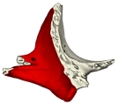 Left zygomatic bone (medial view)
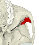 Temporal fossa (from below)
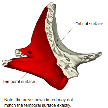
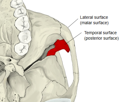
More detail
Anatomy textbooks describe it as follows.
It forms the anterior wall of the temporal fossa and is concave backwards and medially. The zygomaticotemporal foramen opens on it.
Its upper part looks into the temporal fossa, and its lower part towards the infratemporal fossa. The zygomaticotemporal nerve passes through the zygomaticotemporal foramen, and the temporalis lies just behind the surface in the temporal fossa.
Question
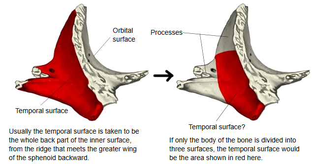
In the left illustration above, the whole back part of the inner surface of the zygomatic bone (except the joint with the temporal bone) is shown in red. The detailed page on the zygomatic bone, however, divides the bone into a 'body' and its processes and says that 'the body has three surfaces', as some anatomy textbooks do. On that view the processes are not part of the surfaces, and the temporal surface is smaller than the red area in the left illustration (see the illustration below). Which view is correct has not been settled here.
Related terms
- Temporal fossa
- Infratemporal crest
- Temporal fascia
- Temporal region
- Zygomatic bone
- Lateral surface of zygomatic bone
- Zygomaticotemporal foramen
- Orbital margin of zygomatic bone
- Temporal process
- Zygomatic arch
- Infratemporal fossa
References and sources
- Further reading: Zygomatic bone, Wikipedia.
- Federative International Programme for Anatomical Terminology (FIPAT). Terminologia Anatomica, 2nd ed. (TA2), 2019.