Medial condyle of tibia (Latin: condylus medialis tibiae)
The part that projects on the medial side of the upper end of the tibia (the end that articulates with the femur). Its upper surface is the superior articular surface, covered with cartilage. The femur has a part with the same name, the medial condyle of femur. The projection on the lateral side is called the lateral condyle.
· Of the two condyles of the upper end of the tibia, the medial condyle is the larger. Its articular surface is oval and slightly hollow; it carries the medial condyle of the femur, with the medial meniscus between them, and is separated from the articular surface of the lateral condyle by the intercondylar eminence.
· On the back of the condyle a horizontal groove receives the main insertion of semimembranosus. The tibial collateral ligament attaches to its medial margin and to the medial surface of the tibia below it.
· Unlike the lateral condyle, it has no facet for the fibula. It forms part of the proximal epiphysis of the tibia and of the knee joint.
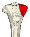 Right tibia, upper end (anterior)
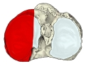 Right tibia (superior)
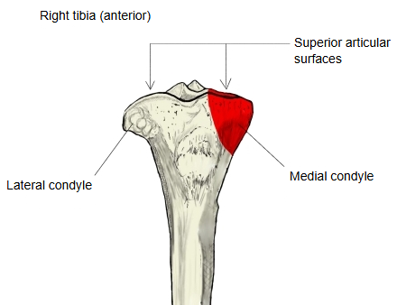
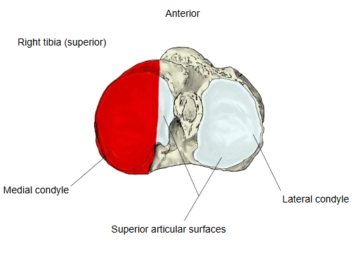
Attachments
| Muscle | Semimembranosus (insertion) | musculus semimembranosus |
| Ligament | Tibial collateral ligament (medial collateral ligament) | ligamentum collaterale tibiale |
Reference websites
[Wikipedia]
· Medial condyle of tibia, Wikipedia.
· Tibia, Wikipedia.
[Websites with illustrations]
· Upper surface of the right tibia (Gray's Anatomy, fig. 257) (Wikimedia Commons)

· Bones of the right leg, anterior surface (Gray's Anatomy, fig. 258) (Wikimedia Commons)

· The tibia (TeachMeAnatomy)
Where it is found
Related terms
- Lateral condyle of tibia
- Superior articular surface of tibia
- Intercondylar eminence
- Proximal epiphysis of tibia
- Medial condyle of femur
- Knee joint
- Tibia
References and sources
- Further reading: Medial condyle of tibia, Wikipedia.
- Further reading: Tibia, Wikipedia.
- Federative International Programme for Anatomical Terminology (FIPAT). Terminologia Anatomica, 2nd ed. (TA2), 2019.