Cribriform plate (Latin: lamina cribrosa ossis ethmoidalis)
The cribriform plate is one of the three parts of the ethmoid bone: the thin horizontal plate in the middle.
Ethmoid bone: one of the bones of the neurocranium
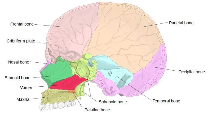
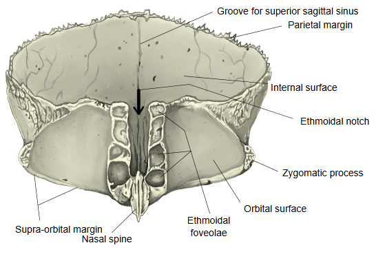
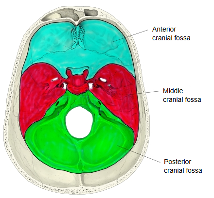
The cribriform plate fits into the ethmoidal notch of the frontal bone, between its two orbital parts, and this thin horizontal plate forms part of the floor of the anterior cranial fossa; below, it is the roof of the nasal cavity.
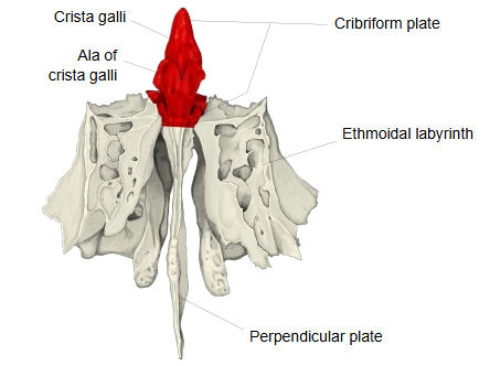
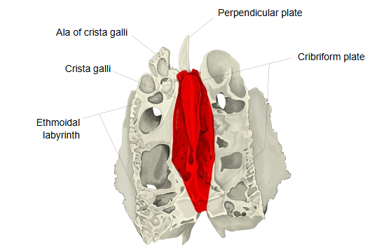
From the midline of the cribriform plate, highest at the front, rises a process called the crista galli; from its anterior border two small processes, the alae (wings) of the crista galli, project forwards.
Because of this process, the cribriform plate seen from the front appears to project upwards.
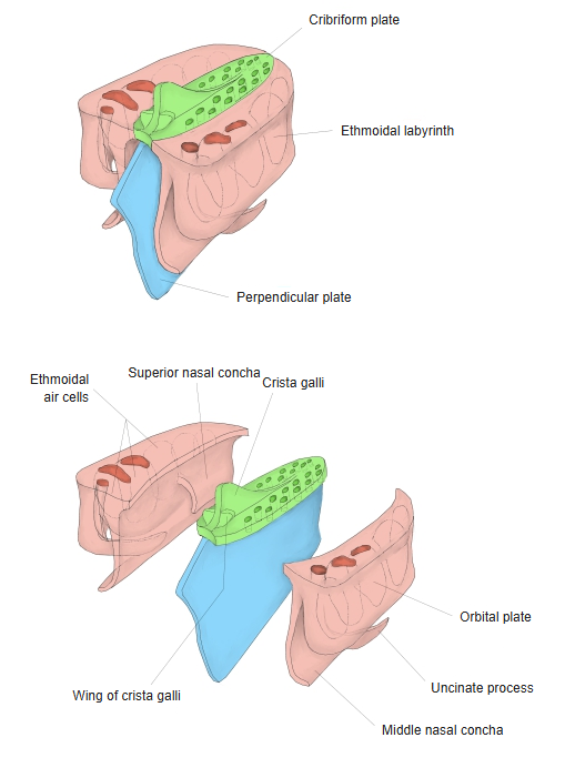
- Cribriform plate : the horizontal bony plate in the middle.
- Perpendicular plate : the plate that projects downwards from the underside of the cribriform plate.
- Ethmoidal labyrinth : the parts on both sides of the perpendicular plate that hang down from the cribriform plate.
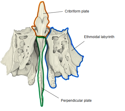
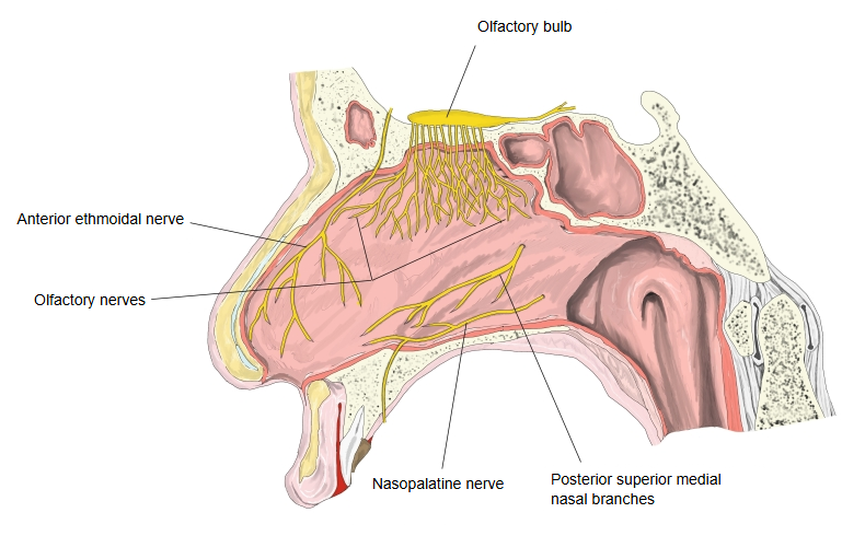
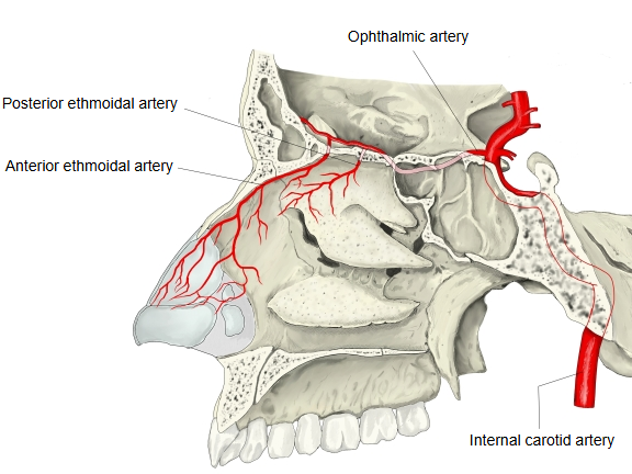
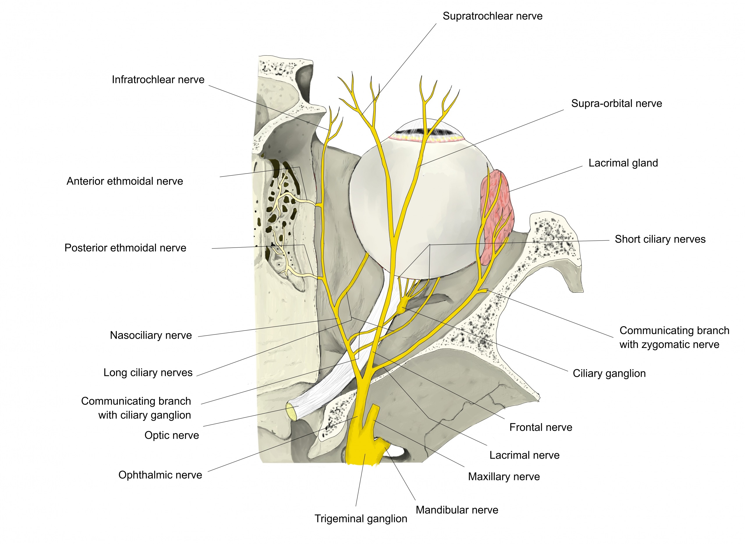
Many small foramina open in the horizontal part of the cribriform plate (not in the crista galli). Through them pass the olfactory nerves, the anterior ethmoidal nerve and the nasal branches of the anterior and posterior ethmoidal arteries and ethmoidal veins.
On each side of the crista galli the plate forms a narrow groove that holds the olfactory bulb. The olfactory nerve fibres pass through its foramina to the roof of the nasal cavity, the upper part of the nasal septum and the superior nasal concha. Near its front a slit on each side admits a fold of dura mater, and a small notch lets the anterior ethmoidal nerve and vessels enter the nose.
A fracture of the cribriform plate can tear the olfactory nerves (loss of smell) and the dura, so that cerebrospinal fluid leaks from the nose (CSF rhinorrhoea), with a risk of meningitis.
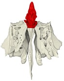 Ethmoid bone (anterior)
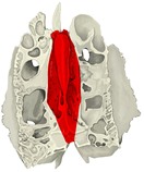 Ethmoid bone (superior)
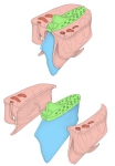 Parts of the ethmoid bone
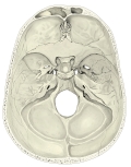 Internal surface of the cranial base
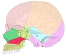 Skull (sagittal section, bones coloured)
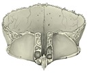 Frontal bone (inferior)
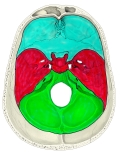 Cranial fossae
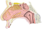 Nerves of the nasal septum
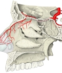 Ethmoidal arteries
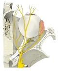 Nerves of the orbit (from above)
Reference websites
[Wikipedia]
· Cribriform plate, Wikipedia.
· Ethmoid bone, Wikipedia.
Where it is found
Related terms
- Crista galli
- Perpendicular plate
- Ethmoidal labyrinth
- Ethmoid bone
- Orbital plate
- Ethmoidal air cells
- Nasal cavity
References and sources
- Further reading: Cribriform plate, Wikipedia.
- Further reading: Ethmoid bone, Wikipedia.
- Federative International Programme for Anatomical Terminology (FIPAT). Terminologia Anatomica, 2nd ed. (TA2), 2019.