Orbital plate (Latin: lamina orbitalis)

Also called Orbital plate of ethmoid or the lamina papyracea
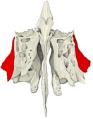
The orbital plate is the smooth, thin plate of bone that forms the lateral surface of the ethmoidal labyrinth of the ethmoid bone; it forms the largest part of the medial wall of the orbit.
Ethmoidal labyrinth: one of the main parts of the ethmoid bone, together with the cribriform plate and the perpendicular plate
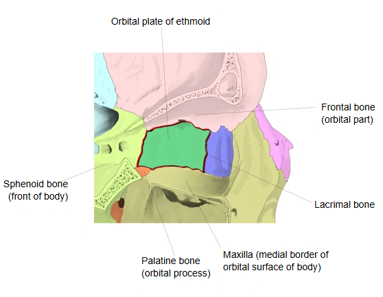
Its four borders and the bones they meet: front border → lacrimal bone; back border → sphenoid bone; upper border → frontal bone (orbital part); lower border → maxilla, and at its back end the orbital process of the palatine bone.
Sphenoid bone: the front of the body of the sphenoidMaxilla: the medial edge of the orbital surface of the body of the maxilla
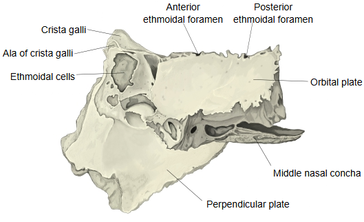
Where its upper border joins the orbital part of the frontal bone, the two bones form the anterior and posterior ethmoidal foramina, which carry nerves and vessels.
Nerves and vessels: the anterior and posterior ethmoidal nerves, the anterior and posterior ethmoidal arteries, and the anterior and posterior ethmoidal veins
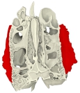
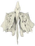
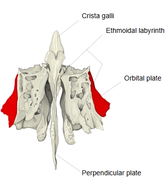
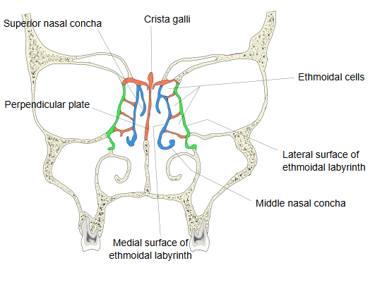
  • The ethmoid bone from the front, with the orbital plates in red on the outer sides of the labyrinths.Ethmoid bone (anterior)
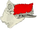
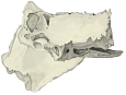
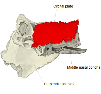
  • The ethmoid bone from the left side, with the orbital plate in red.Ethmoid bone (left lateral)
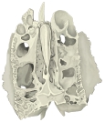
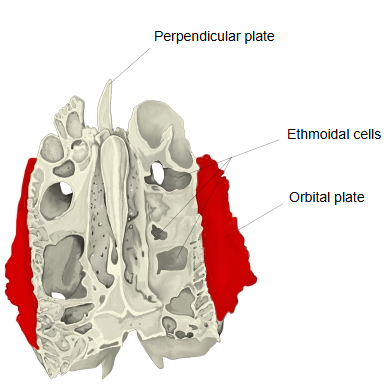
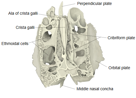
  • The ethmoid bone from above, with the orbital plates in red along its sides.Ethmoid bone (superior)
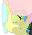
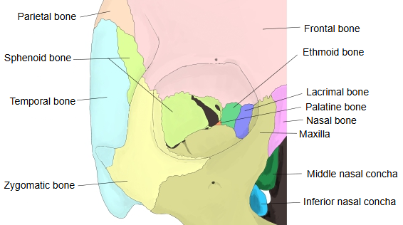
  • The right orbit from the front with each bone in a different colour, the ethmoid in green on the medial wall.Bones of the right orbit
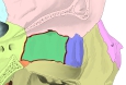
  • The medial wall of the right orbit with each bone coloured, the orbital plate in green outlined in red with the bones that border it.Medial wall of the right orbit
  • The ethmoid bone from the front, showing the crista galli, ethmoidal cells, orbital plate, nasal conchae and perpendicular plate.Ethmoid bone (anterior, labelled)
  • The ethmoid bone from the left side, with the orbital plate and the anterior and posterior ethmoidal foramina along its upper edge.Ethmoid bone (left lateral, labelled)
  • The ethmoid bone from above, with the cribriform plate in the middle and the orbital plates along the sides.Ethmoid bone (superior, labelled)
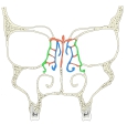
  • Diagram of a coronal section through the orbits and nasal cavity; the green line on each side is the orbital plate between the orbit and the ethmoidal cells.Coronal section through the orbits
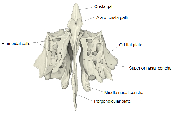
It is the outer wall of the ethmoidal labyrinth and covers the middle and posterior ethmoidal cells; it is rectangular and smooth.

Below the plate, the labyrinth has a surface that joins the palatine bone and the body of the maxilla; some ethmoidal cells open there as well and are closed by these bones.

· It is paper-thin, hence its old name lamina papyracea. It is not to be confused with the orbital part of the frontal bone, which some older texts also call an orbital plate.

· Because it is so thin, it fractures easily in blows to the orbit (medial blow-out fracture), sometimes trapping the medial rectus muscle; infection can also pass through it from the ethmoidal cells to the orbit.

Reference websites

[Wikipedia]

· Orbital lamina of ethmoid bone, Wikipedia.

· Orbit (anatomy), Wikipedia.

[Websites with illustrations]

· Ethmoid bone (Kenhub)

· The Bony Orbit (TeachMeAnatomy)

Where it is found

References and sources

  1. Further reading: Orbital lamina of ethmoid bone, Wikipedia.
  2. Further reading: Orbit (anatomy), Wikipedia.
  3. Federative International Programme for Anatomical Terminology (FIPAT). Terminologia Anatomica, 2nd ed. (TA2), 2019.