Ethmoidal labyrinth (Latin: labyrinthus ethmoidalis)
Also called: lateral mass of ethmoid
- Cribriform plate : the horizontal plate in the middle (in the floor of the cranial cavity).
- Perpendicular plate : the plate that projects downwards from the underside of the cribriform plate (in the nasal septum).
- Ethmoidal labyrinth : the parts on each side of the perpendicular plate that hang down from the cribriform plate.
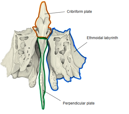
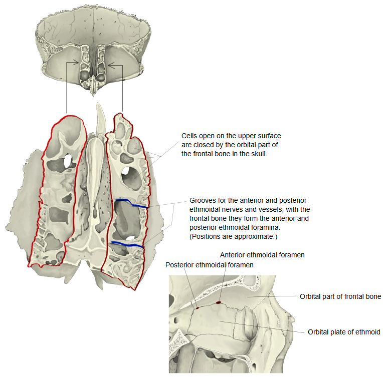
Each labyrinth is made of very thin plates of bone enclosing the ethmoidal cells, arranged in anterior, middle and posterior groups. Many cells are open in the isolated bone; in the skull they are closed by the neighbouring bones.
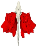 Ethmoid bone (anterior)
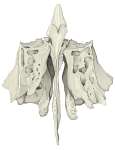 Ethmoid bone (anterior, labelled)
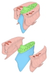 Parts of the ethmoid bone
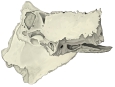 Ethmoid bone (left lateral)
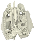 Ethmoid bone (superior)
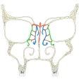 Coronal section through the orbits
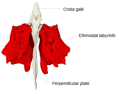
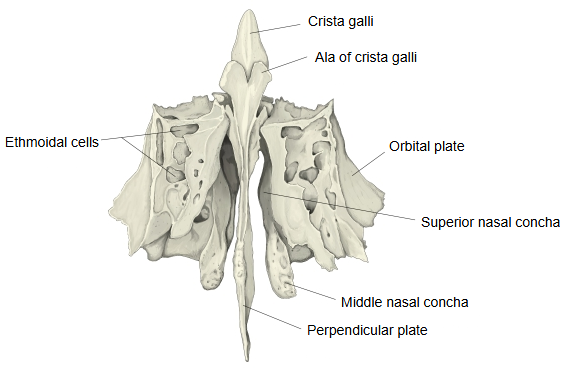
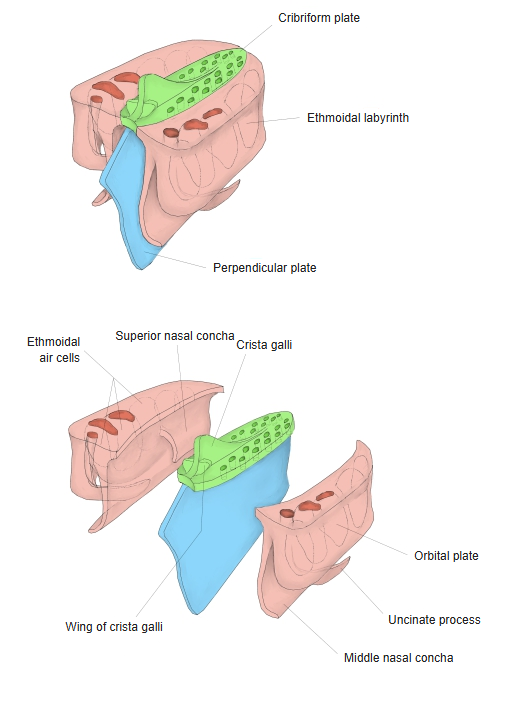
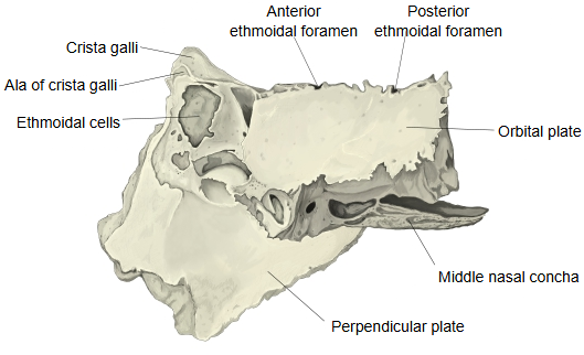
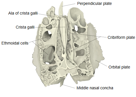
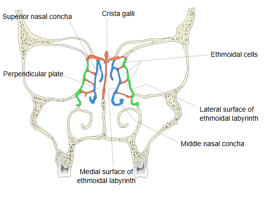
Main surfaces
· Upper surface: many ethmoidal cells lie open here; they are completed by the edges of the ethmoidal notch of the frontal bone, with which this surface joins. Two grooves across it become the anterior and posterior ethmoidal foramina when the frontal bone is in place; they open on the medial wall of the orbit.
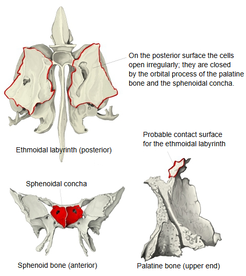
· Posterior surface: large, irregular ethmoidal cells lie open here; they are closed by the sphenoidal concha and the orbital process of the palatine bone, with which this surface joins.
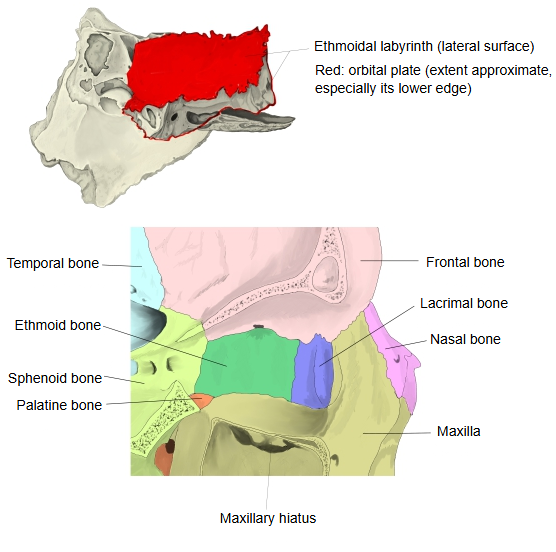
· Lateral surface: the thin, smooth, oblong orbital plate, which forms a large part of the medial wall of the orbit.
In front of the orbital plate, broken cells are closed by the lacrimal bone and the frontal process of the maxilla, and the curved uncinate process runs downwards and backwards from the front part of the labyrinth.
Joints of the orbital plate: above, the frontal bone (orbital part); below, the maxilla and the palatine bone (orbital process); in front, the lacrimal bone; behind, the sphenoid bone.
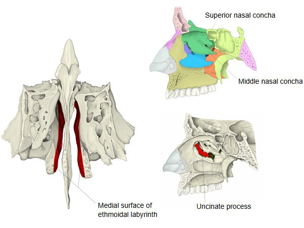
· Medial surface: part of the lateral wall of the nasal cavity. It carries the superior nasal concha and the middle nasal concha, with the superior nasal meatus between them; its upper part is grooved by branches of the olfactory nerve.
Openings of the ethmoidal cells
The posterior cells open into the superior nasal meatus.
The middle cells open into the middle nasal meatus, often on the ethmoidal bulla.
The anterior cells open into the middle nasal meatus through the ethmoidal infundibulum, which in some skulls also continues up to the frontal sinus.
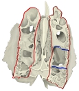 Upper surface of the labyrinth
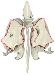 Posterior surface of the labyrinth
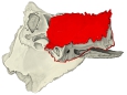 Lateral surface of the labyrinth
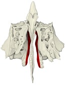 Medial surface of the labyrinth
Reference websites
[Wikipedia]
· Ethmoidal labyrinth, Wikipedia.
· Ethmoid bone, Wikipedia.
[Websites with illustrations]
· Ethmoid bone: anatomy, borders and development (Kenhub)
· Ethmoid bone from above (Gray's Anatomy, fig. 149) (Wikimedia Commons)

· Ethmoid bone from behind (Gray's Anatomy, fig. 151) (Wikimedia Commons)

· Ethmoid bone from the right side (Gray's Anatomy, fig. 152) (Wikimedia Commons)

Where it is found
Related terms
- Ethmoidal air cells
- Orbital plate
- Cribriform plate
- Perpendicular plate
- Superior nasal concha
- Middle nasal concha
- Ethmoid bone
References and sources
- Further reading: Ethmoidal labyrinth, Wikipedia.
- Further reading: Ethmoid bone, Wikipedia.
- Federative International Programme for Anatomical Terminology (FIPAT). Terminologia Anatomica, 2nd ed. (TA2), 2019.