Ethmoidal air cells (Latin: cellulae ethmoidales)
Also called Ethmoidal cells or the ethmoidal sinus
· The term means the same as the ethmoidal sinus.
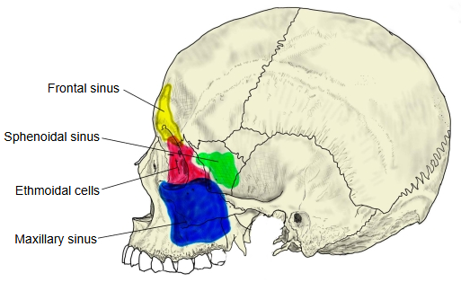
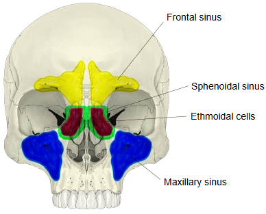
- Maxillary sinus : in the maxilla; the largest of the paranasal sinuses
- Frontal sinus : in the frontal bone
- Ethmoidal sinus : in the ethmoid bone; divided into many separate spaces (the ethmoidal air cells)
- Sphenoidal sinus : in the sphenoid bone, lying behind the ethmoidal sinus
· There are no sharp borders between the groups. The number and size of the cells vary widely between people, and cells that spread into neighbouring bones are named after their position, for example infra-orbital (Haller) cells in the floor of the orbit.
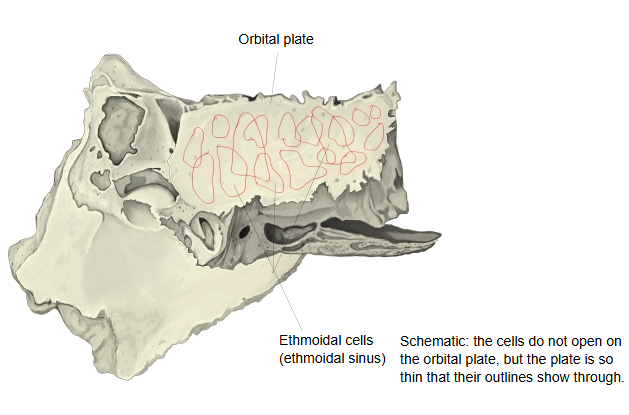
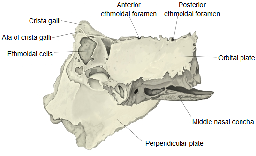
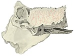 Ethmoid bone (left lateral, cells outlined)
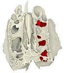 Ethmoid bone (superior, openings)
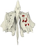 Ethmoid bone (anterior, openings)
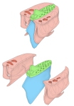 Parts of the ethmoid bone
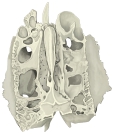 Ethmoid bone (superior)
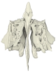 Ethmoid bone (anterior)
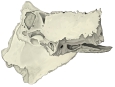 Ethmoid bone (left lateral)
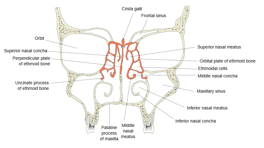
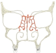 Nasal cavity (coronal section)
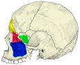 Paranasal sinuses (lateral)
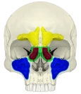 Paranasal sinuses (anterior)
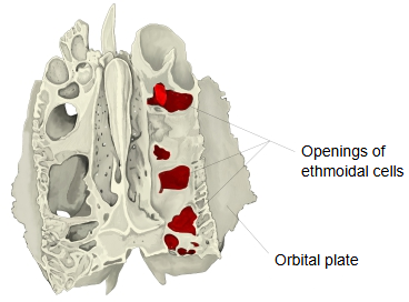
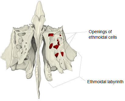
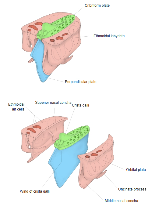
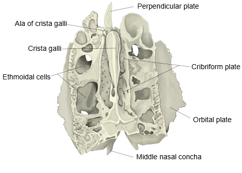
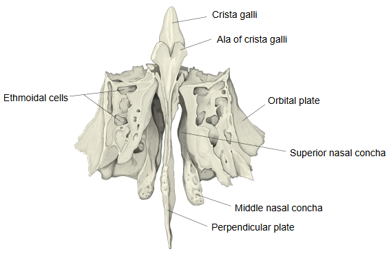
Openings
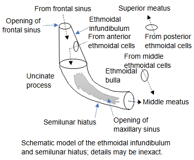
- Anterior cells : open into the ethmoidal infundibulum and through the semilunar hiatus into the middle nasal meatus
- Middle cells : open on or above the ethmoidal bulla into the middle nasal meatus
- Posterior cells : open into the superior nasal meatus; occasionally one opens into the sphenoidal sinus
Neighbouring bones
- Above : the ethmoidal foveolae on the orbital part of the frontal bone
- In front : the nasal part of the frontal bone, the frontal process of the maxilla and the lacrimal bone
- Below : related to the nasal cavity and the maxilla
- Behind : the sphenoidal concha and the orbital process of the palatine bone
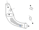 Ethmoidal infundibulum and semilunar hiatus (model)
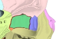 Medial wall of the right orbit
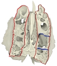 Upper surface of the ethmoid
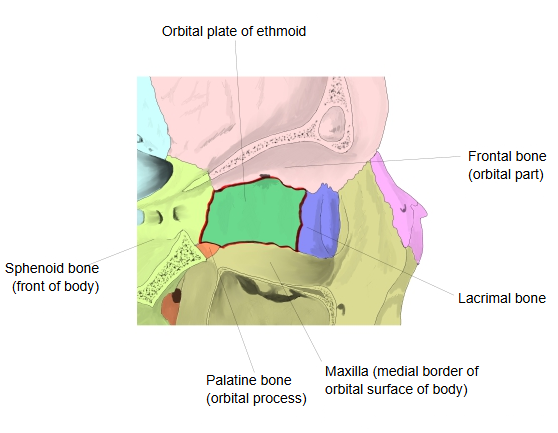
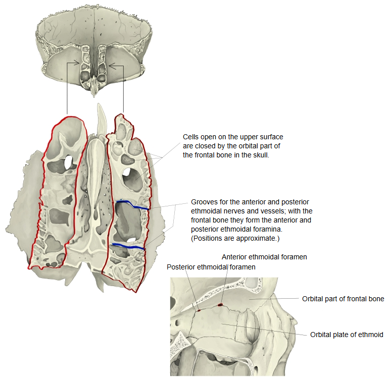
The cells lie between the nasal cavity and the orbit. They are separated from the orbit only by the paper-thin orbital plate, and from the nasal cavity by the thin medial wall of the labyrinth. Like the other paranasal sinuses they are lined with mucous membrane continuous with that of the nose. They are present at birth, together with the maxillary sinuses, and cannot be felt from outside.
Nerve supply: sensory fibres come from the anterior and posterior ethmoidal nerves (from the nasociliary nerve) and from orbital branches of the pterygopalatine ganglion. Secretory (parasympathetic) fibres from the facial nerve reach the lining through the pterygopalatine ganglion.
Because the orbital plate is so thin, infection of the ethmoidal cells (ethmoiditis) can spread into the orbit.
Reference websites
[Wikipedia]
· Ethmoid sinus, Wikipedia.
· Paranasal sinuses, Wikipedia.
[Websites with illustrations]
· The paranasal sinuses (TeachMeAnatomy)
· Ethmoid bone from behind, with the labyrinths (Gray's Anatomy) (Wikimedia Commons)

Where it is found
Related terms
References and sources
- Further reading: Ethmoid sinus, Wikipedia.
- Further reading: Paranasal sinuses, Wikipedia.
- Federative International Programme for Anatomical Terminology (FIPAT). Terminologia Anatomica, 2nd ed. (TA2), 2019.