Latin: nervus femoralis
Overview
- The largest branch of the lumbar plexus.
- Mixed nerve: motor to the front of the thigh, sensory to the skin of the front of the thigh and the medial side of the leg and foot.
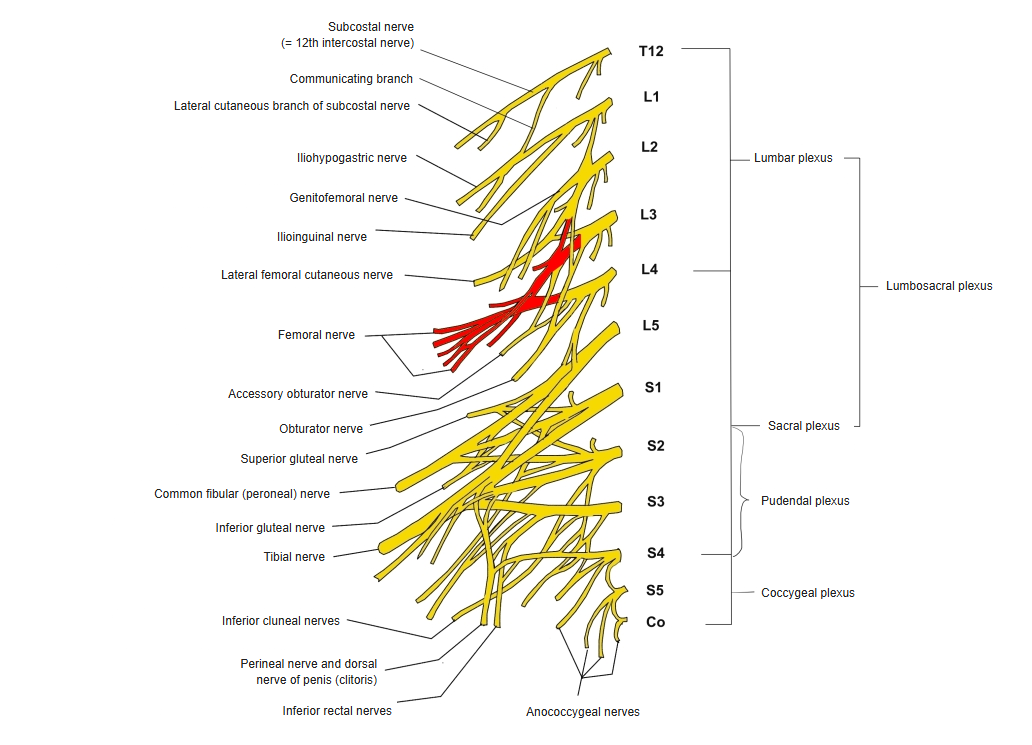
- Formed from the ventral rami of L2–L4.
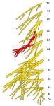
Lumbar and sacral plexuses 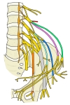
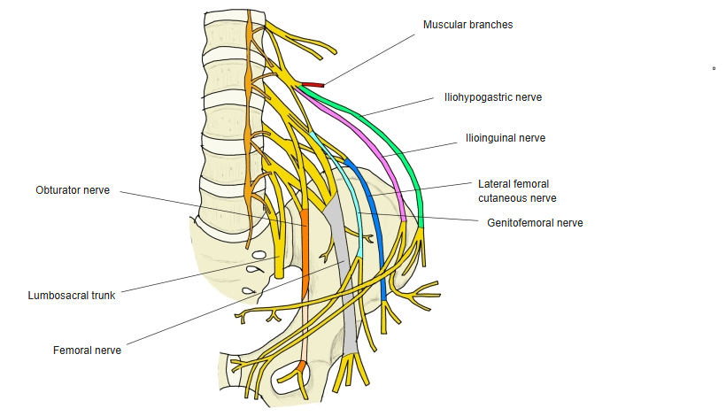
Lumbar plexus and its branches
Course
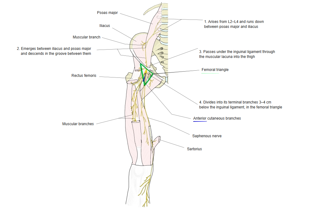
- Descends in the groove between psoas major and iliacus, giving branches to both.
- Passes under the inguinal ligament through the muscular lacuna into the thigh, lateral to the femoral artery.
- Divides into its terminal branches in the femoral triangle, about 3–4 cm below the inguinal ligament.
Course of the femoral nerve 
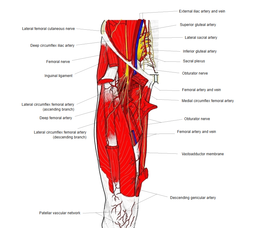
Femoral nerve and nearby vessels
Branches and supply
| Branch | Supplies |
|---|---|
| Muscular branches | Iliacus, psoas major, pectineus, sartorius and the quadriceps femoris (rectus femoris, vastus medialis, vastus intermedius, vastus lateralis); articularis genus |
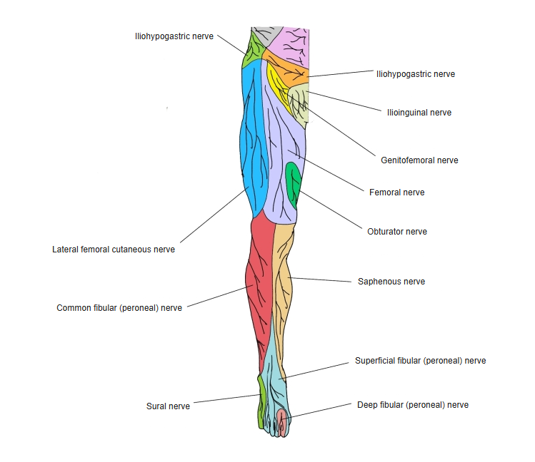
| Anterior cutaneous branches | Skin of the front of the thigh and along the medial border of sartorius |
| Saphenous nerve | Skin below the knee on the medial side, the medial side of the leg and the medial border of the foot |
| Articular branches | Hip joint and knee joint |
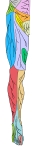
Cutaneous nerves of the lower limb (front) 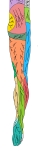
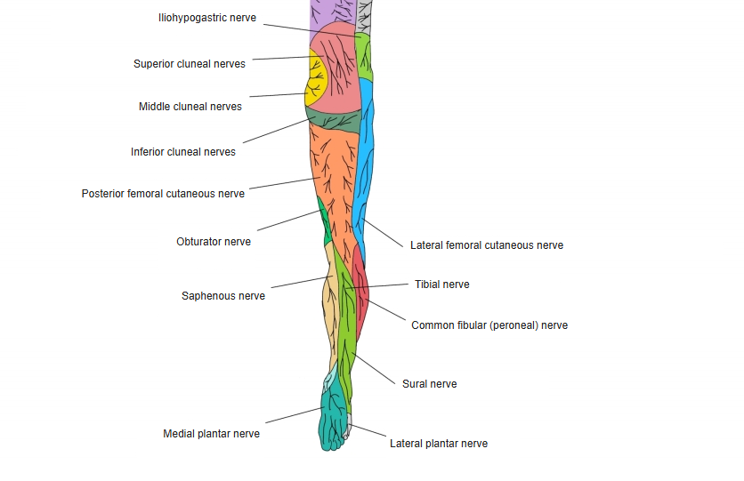
Cutaneous nerves of the lower limb (back)
References and sources
- Further reading: Femoral nerve, Wikipedia.