Latin: cornea
Overview
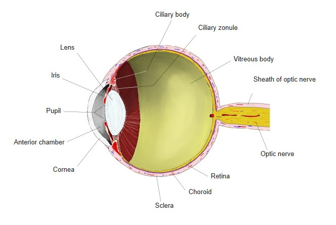
- The transparent front part of the fibrous layer, covering the iris, pupil and anterior chamber.
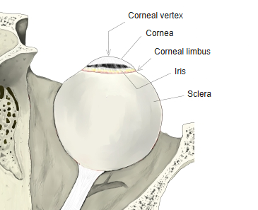
- Occupies about one-sixth of the eyeball surface and bulges forward slightly more than the sclera; the two meet at the corneal limbus.
- The main focusing surface of the eye: about two-thirds of its refractive power (around 43 dioptres); its focus is fixed – fine focusing is done by the lens.
- Has no blood vessels: it gets oxygen from the tear film and nutrients from the aqueous humor and the vessels at the limbus.
- Very richly supplied with sensory nerve endings; touching it triggers the blink (corneal) reflex.
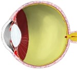
Eyeball, longitudinal section 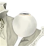
Front of the eyeball
Shape and size
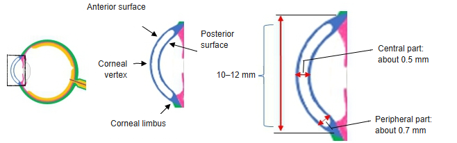
- Horizontal diameter about 11.5–12 mm, vertical about 10.5–11 mm.
- Thickness about 0.5–0.6 mm in the centre and up to about 0.7 mm or more at the periphery.
- The posterior surface is more strongly curved than the anterior surface.
- The most forward point is the corneal vertex; refractive index about 1.376.
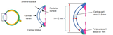
Cornea: surfaces and thickness
Layers
| Layer (front to back) | Features |
|---|---|
| Corneal epithelium | Non-keratinized stratified squamous epithelium, about 50–60 µm; continuous with the conjunctival epithelium at the limbus; regenerates quickly and contains free nerve endings |
| Anterior limiting lamina (Bowman's layer) | Acellular layer of fine collagen fibrils, about 10–20 µm; fades out towards the limbus |
| Substantia propria (stroma) | About 90% of the thickness; regular sheets of collagen fibrils at right angles to each other, with flattened keratocytes – the regular arrangement makes it transparent |
| Posterior limiting lamina (Descemet's membrane) | Basement membrane of the endothelium, about 6–10 µm, thickening with age; separates easily |
| Corneal endothelium | Single layer of flat cells; pumps fluid out of the stroma and so keeps it clear |
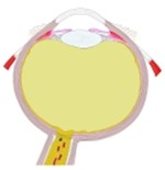
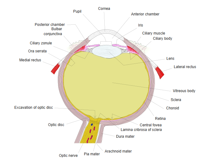
Right eyeball, horizontal section (model)
References and sources
- Further reading: Cornea, Wikipedia.