Carotid sheath (Latin: vagina carotica)
The carotid sheath is
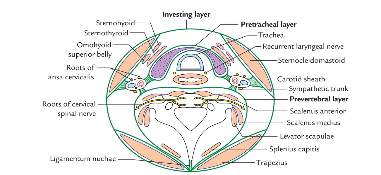
- Investing (superficial) layer : the outermost layer; it surrounds the whole neck, but may be incomplete in places.
- Pretracheal layer : encloses the organs around the trachea and the muscles at the front of the neck.
- Prevertebral layer : encloses the cervical vertebrae and the muscles around them.
- Carotid sheath : encloses the common carotid artery, the internal jugular vein, the vagus nerve and other structures.
The carotid sheath mainly encloses the following four structures.
1. 1. Common carotid artery: the main artery to the head; it divides into the internal and external carotid arteries. Above the bifurcation the sheath contains the internal carotid artery; it lies medially in the sheath.
2. 2. Internal jugular vein: the main vein that returns the blood of the brain to the heart; it lies laterally in the sheath.
3. 3. Vagus nerve: the tenth cranial nerve, a mixed nerve distributed widely to the organs of the neck, chest and abdomen; it lies behind, in the groove between the artery and the vein.
4. 4. Deep cervical lymph nodes: along the internal jugular vein.
In addition:
In its upper part, the glossopharyngeal, accessory and hypoglossal nerves pass briefly through the sheath, piercing its fascia.
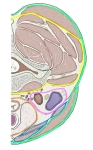 Cervical fascia (C6 level)
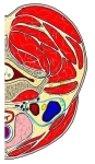 Carotid sheath
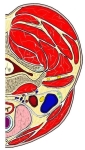 Cross-section at C6
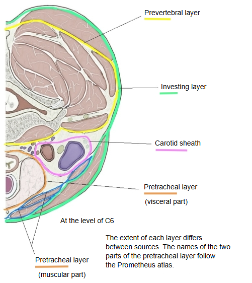
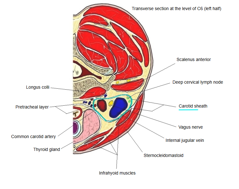
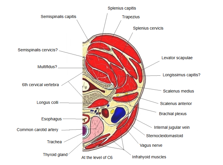
On each side of the neck, the common carotid artery runs upward between the deep surface of the sternocleidomastoid and the prevertebral layer and continues into the internal and external carotid arteries; the internal jugular vein and the vagus nerve run with them. The strong fibrous membrane that wraps this bundle of large vessels and nerves together is a vascular nerve sheath called the carotid sheath. Sources differ on the external carotid artery: most English texts place it outside the sheath; some include it.
It is also described as follows.
The sheath is made of fascia from the pretracheal and investing layers of the cervical fascia, together with fascia joining them to the prevertebral layer. The sympathetic trunk lies just behind the sheath, and the ansa cervicalis is embedded in its front wall. The intermediate tendon of the omohyoid is held by a fascial sling joined to the sheath; contraction of the muscle is thought to tense the sheath and help keep the internal jugular vein open, aiding the return of venous blood from the head.
Reference websites
[Wikipedia]
· Carotid sheath, Wikipedia.
· Deep cervical fascia, Wikipedia.
· Investing layer: encloses the sternocleidomastoid and trapezius.
· Carotid sheath: encloses the vascular compartment of the neck.
· Pretracheal layer: encloses the visceral compartment of the neck (trachea, oesophagus and thyroid gland), and with its muscular part the infrahyoid muscles.
· Prevertebral layer: encloses the vertebral compartment of the neck.
Structure
The carotid sheath lies on each side of the neck, deep to the sternocleidomastoid, at the side of the pharynx and trachea and lateral to the retropharyngeal space (at the level of the oropharynx). It runs from the base of the skull to the root of the neck (the first rib and the sternum).
Contents
Its four main contents are:
· the common carotid artery, and above it the internal carotid artery (medial)
· the internal jugular vein (lateral)
· the vagus nerve (behind, between the two vessels)
· the deep cervical lymph nodes
The artery lies medial to the vein, and the vagus nerve lies behind, between them. In the upper part the glossopharyngeal, accessory and hypoglossal nerves pass through the sheath. The ansa cervicalis, formed by the superior root (fibres of C1 running with the hypoglossal nerve) and the inferior root (C2–C3), is embedded in its front wall.
Layers
All three main layers of the deep cervical fascia (investing, pretracheal and prevertebral) contribute to the carotid sheath.
Useful sites

· Deep cervical fascia (figure, Earth's Lab)
· Carotid sheath (figure, Wikipedia)

Where it is found
Related terms
- Common carotid artery
- Internal carotid artery
- Internal jugular vein
- Vagus nerve
- Ansa cervicalis
- Sympathetic trunk
- Omohyoid
References and sources
- Further reading: Carotid sheath, Wikipedia.
- Further reading: Deep cervical fascia, Wikipedia.
- Federative International Programme for Anatomical Terminology (FIPAT). Terminologia Anatomica, 2nd ed. (TA2), 2019.