Lobe of lung (Latin: lobus pulmonis)
Also called Pulmonary lobe
of the
lungs
- Right lung
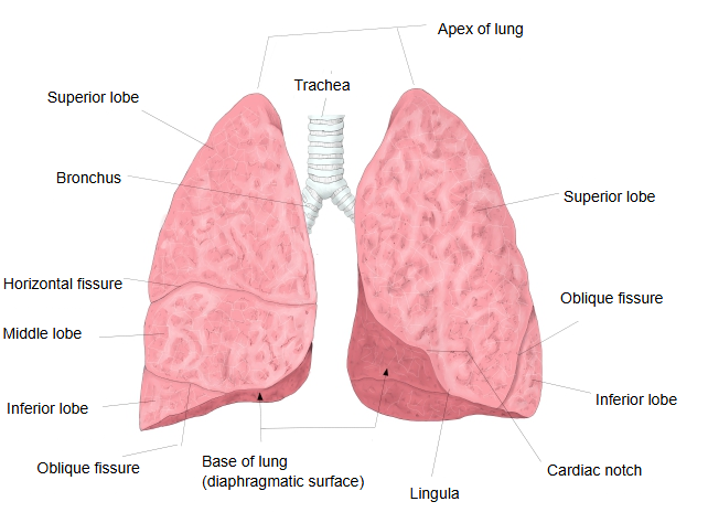
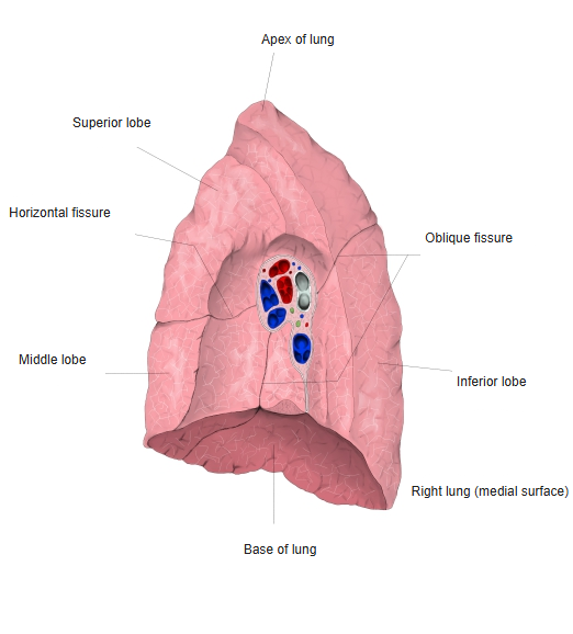
- Superior lobe : its lower edge in front (the horizontal fissure) runs forward roughly along the 4th rib to the 4th costal cartilage; behind, where the oblique fissure begins, it reaches down to about the 4th rib. Segments S1-S3.
- Middle lobe : lies between the 4th and 6th ribs, in front of the midaxillary line. Segments S4-S5.
- Inferior lobe : lies mainly at the back. Its upper boundary runs obliquely down and forward, from the 4th rib beside the vertebral column to where the midclavicular line crosses the 6th rib. Segments S6-S10.
- Left lung
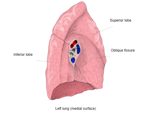
- Superior lobe : its lower end reaches the costochondral junction of the 6th rib; behind, it reaches down to about the 4th rib. Includes the lingula. Segments S1+2, S3, S4 and S5.
- Inferior lobe : segments S6, S7+8 (often counted together), S9 and S10.
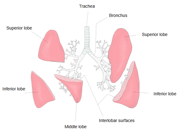
Interlobar surface: the surface where two neighbouring lobes meet.
Pulmonary lobule: a small polygonal area about 1-2 cm across, visible on the surface of each lobe.
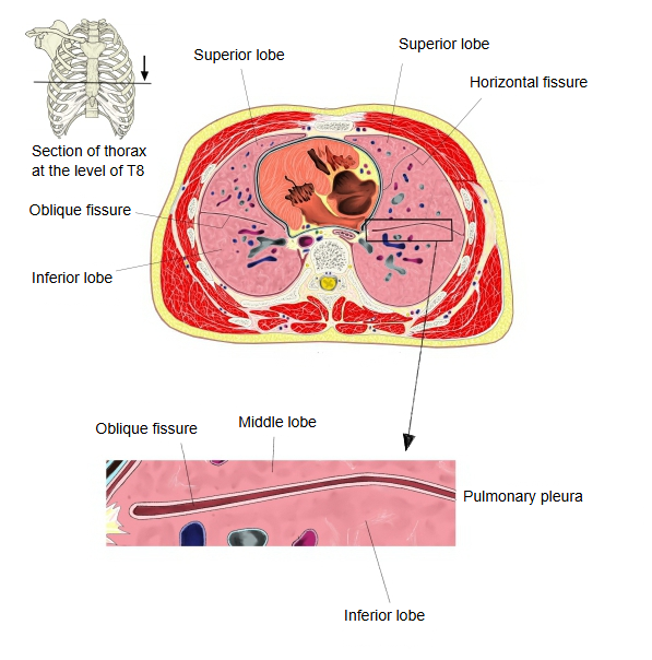
The pulmonary (visceral) pleura covering the lung dips into the oblique and horizontal fissures and separates the lobes almost completely, so that they are joined only at the hilum by their bronchi and vessels; each lobe has its own lobar bronchus. The surfaces where neighbouring lobes meet are the interlobar surfaces. Comparing the lobes, the inferior lobe is shaped like half a cone and is larger than the superior lobe, and the middle lobe is the smallest. On the surface of each lobe, small polygonal areas about 1-2 cm across can be seen with the naked eye; these are the pulmonary lobules. The lobules are bound together by interlobular connective tissue, and where much inhaled dust and carbon is deposited in it, the outlines of the lobules become clearer with age.
The right lung consists of the superior, middle and inferior lobes, from above down; the slightly smaller left lung consists of the superior and inferior lobes, and its lingula corresponds to the right middle lobe. These five lobes are further divided into bronchopulmonary segments: 10 in the right lung and usually 8-10 in the left.
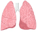 Lungs (anterior view)
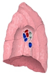 Right lung (medial surface)
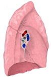 Left lung (medial surface)
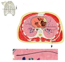 Section of the thorax at T8
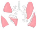 Lobes and interlobar surfaces
Reference websites
[Wikipedia]
· Lobes of lung, Wikipedia.
· Bronchopulmonary segment, Wikipedia.
[Websites with illustrations]
· Lobes of the lungs (figure) (Semantic Scholar)

Related terms
References and sources
- Further reading: Lobes of lung, Wikipedia.
- Federative International Programme for Anatomical Terminology (FIPAT). Terminologia Anatomica, 2nd ed. (TA2), 2019.